BioTalentum Ltd., H-2100 Gödöllő, Hungary.
Department of Anatomy, Cell and Developmental Biology, Eötvös Loránd University, H-1117 Budapest, Hungary.
Cells. 2020 May 1;9(5):1122. doi: 10.3390/cells9051122.
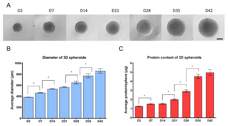
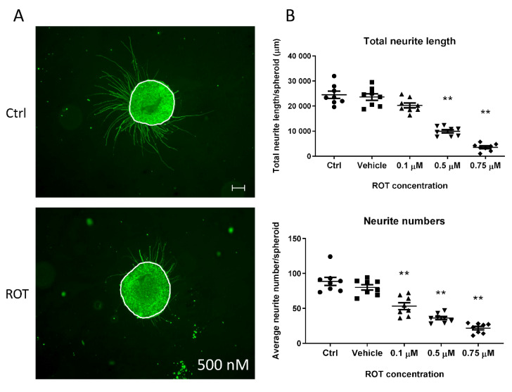
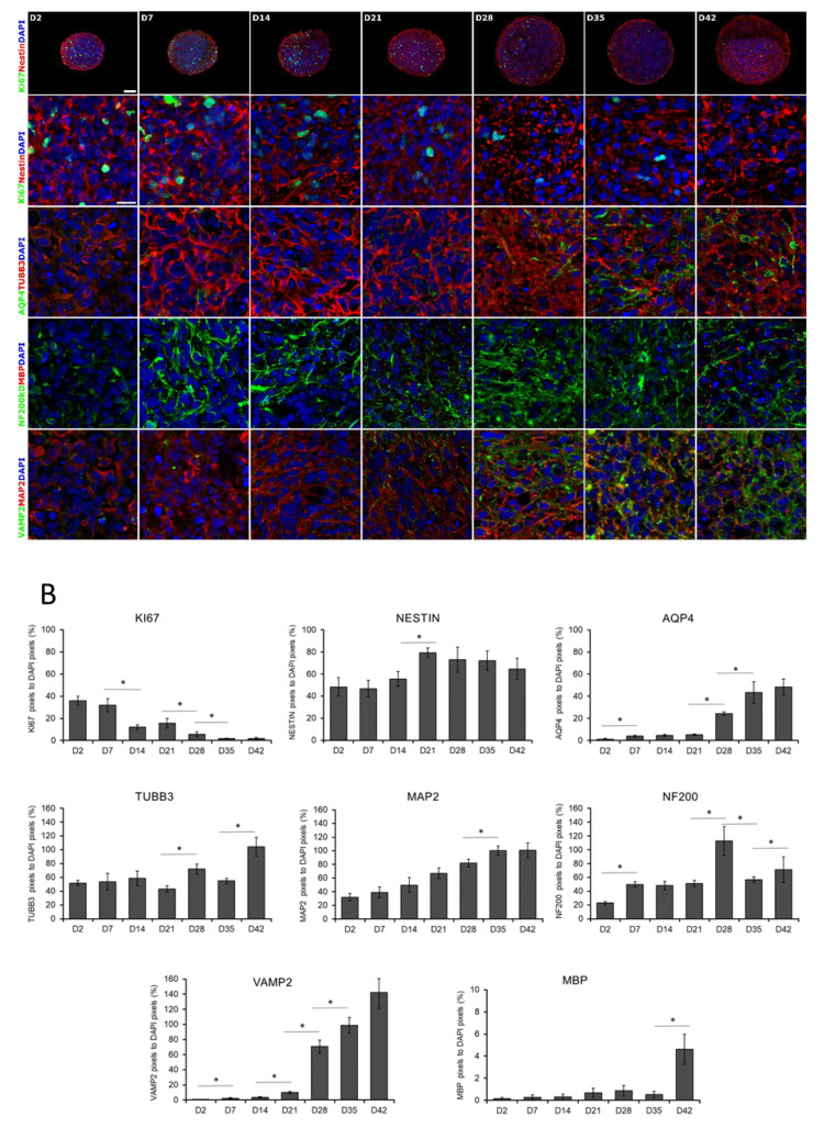
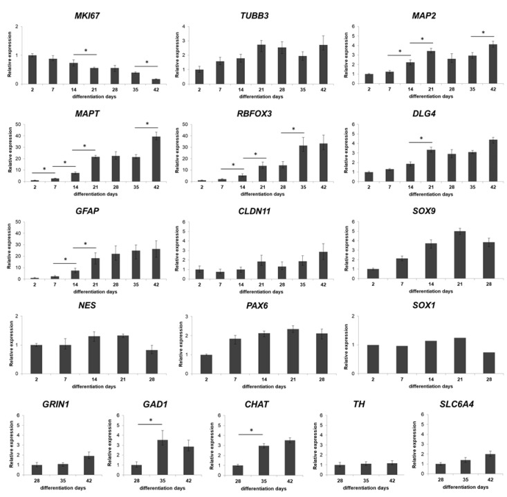
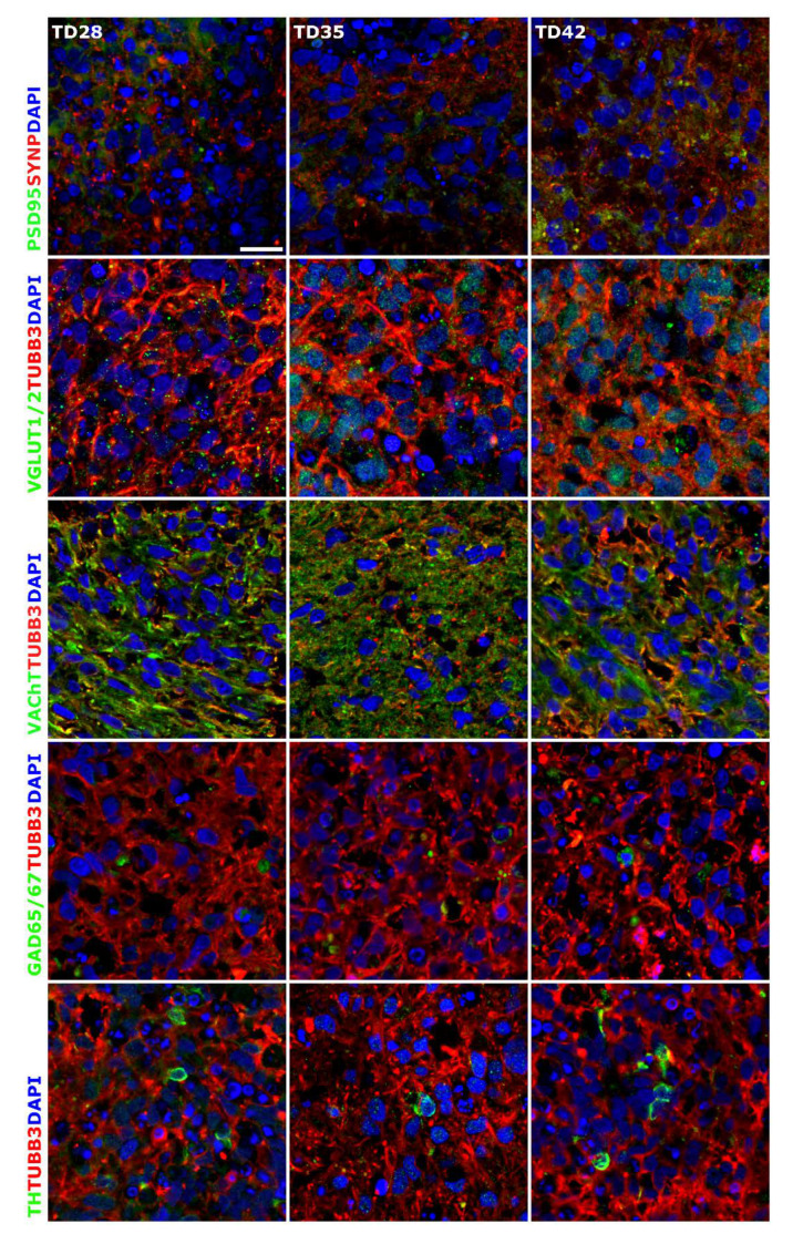
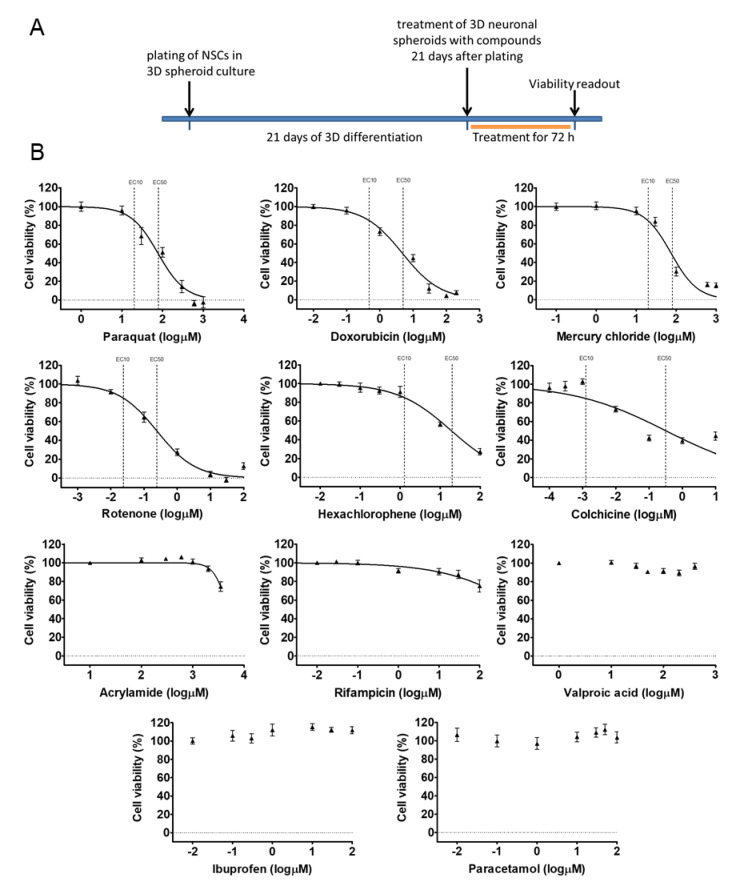
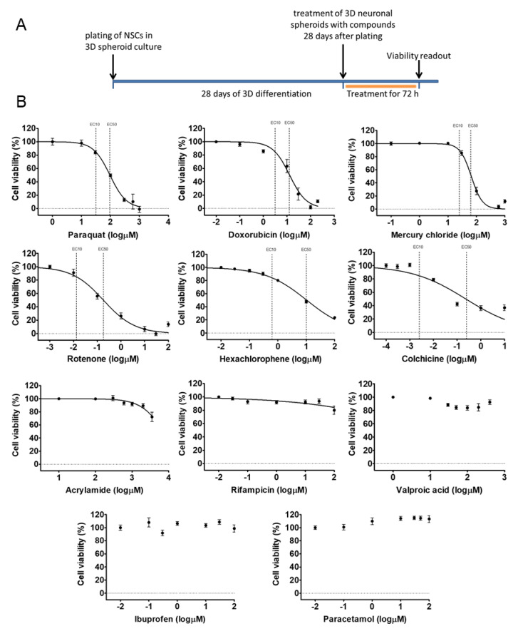
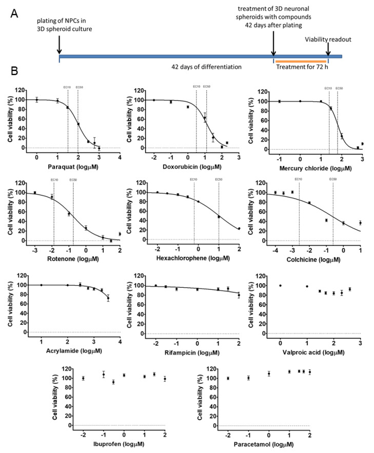
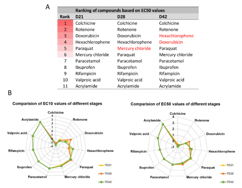
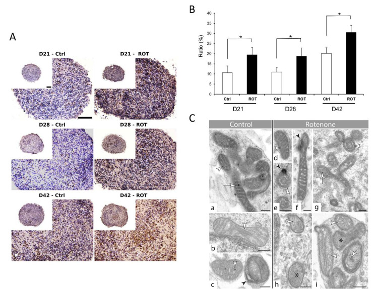
We present a hiPSC-based 3D in vitro system suitable to test neurotoxicity (NT). Human iPSCs-derived 3D neurospheres grown in 96-well plate format were characterized timewise for 6-weeks. Changes in complexity and homogeneity were followed by immunocytochemistry and transmission electron microscopy. Transcriptional activity of major developmental, structural, and cell-type-specific markers was investigated at weekly intervals to present the differentiation of neurons, astrocytes, and oligodendrocytes. Neurospheres were exposed to different well-known toxicants with or without neurotoxic effect (e.g., paraquat, acrylamide, or ibuprofen) and examined at various stages of the differentiation with an ATP-based cell viability assay optimized for 3D-tissues. Concentration responses were investigated after acute (72 h) exposure. Moreover, the compound-specific effect of rotenone was investigated by a panel of ER-stress assay, TUNEL assay, immunocytochemistry, electron microscopy, and in 3D-spheroid based neurite outgrowth assay. The acute exposure to different classes of toxicants revealed distinct susceptibility profiles in a differentiation stage-dependent manner, indicating that hiPSC-based 3D in vitro neurosphere models could be used effectively to evaluate NT, and can be developed further to detect developmental neurotoxicity (DNT) and thus replace or complement the use of animal models in various basic research and pharmaceutical applications.
我们提出了一种基于 hiPSC 的适用于测试神经毒性(NT)的 3D 体外系统。人类 iPSC 来源的 3D 神经球在 96 孔板格式中生长,在 6 周内进行了时间特征分析。通过免疫细胞化学和透射电子显微镜观察复杂性和均一性的变化。每周间隔一次检测主要发育、结构和细胞类型特异性标志物的转录活性,以展示神经元、星形胶质细胞和少突胶质细胞的分化。将神经球暴露于不同的已知有毒物质(如百草枯、丙烯酰胺或布洛芬)中,在分化的不同阶段进行检测,采用优化的基于 ATP 的细胞活力测定法用于 3D 组织。急性(72 h)暴露后进行浓度反应研究。此外,通过一组内质网应激测定、TUNEL 测定、免疫细胞化学、电子显微镜以及基于 3D 球体的神经突生长测定,研究了鱼藤酮的化合物特异性效应。急性暴露于不同类别的有毒物质显示出与分化阶段相关的不同敏感性谱,表明基于 hiPSC 的 3D 体外神经球模型可有效用于评估 NT,并且可以进一步开发以检测发育性神经毒性(DNT),从而在各种基础研究和药物应用中替代或补充动物模型的使用。