Institute of Anatomy, University of Leipzig, Liebigstraße 13, D-04103, Leipzig, Germany.
Institute of Legal Medicine, University of Leipzig, Johannisallee 28, D-04103, Leipzig, Germany.
Int J Legal Med. 2020 Nov;134(6):2187-2193. doi: 10.1007/s00414-020-02308-x. Epub 2020 May 5.
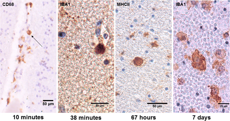
Traumatic brain injury is among the leading causes of death in individuals under 45 years of age. However, since trauma mechanisms and survival times differ enormously, the exact mechanisms leading to the primary and secondary injury and eventually to death after traumatic brain injury (TBI) remain unclear. Several studies showed the versatile functions of microglia, the innate macrophages of the brain, following a TBI. Earlier being characterized as rather neurotoxic, neuroprotective capacities were recently demonstrated, therefore, making microglia one of the key players following TBI. Especially in cases with only short survival times, immediate microglial reactions are of great forensic interest in questions of wound age estimation. Using standardized immunohistochemical methods, we examined 8 cases which died causatively of TBI with survival times between minutes and 7 days and 5 control cases with cardiovascular failure as the cause of death to determine acute changes in microglial morphology and antigen expression after TBI. In this pilot study, we detected highly localized changes in microglial morphology already early after traumatic damage, e.g., activated microglia and phagocyted erythrocytes in the contusion areas in cases with minute survival. Furthermore, an altered antigen expression was observed with increasing trauma wound age, showing similar effects like earlier transcriptomic studies. There is minute data on the direct impact of shear forces on microglial morphology. We were able to show localization-depending effects on microglial morphology causing localized dystrophy and adjacent activation. While rodent studies are widespread, they fail to mimic the exact mechanisms in human TBI response. Therefore, more studies focusing on cadaveric samples need to follow to thoroughly define the mechanisms leading to cell destruction and eventually evaluate their forensic value.
创伤性脑损伤是 45 岁以下人群死亡的主要原因之一。然而,由于创伤机制和存活时间差异巨大,导致创伤性脑损伤(TBI)后原发性和继发性损伤以及最终死亡的确切机制仍不清楚。几项研究表明,TBI 后小胶质细胞(大脑的固有巨噬细胞)具有多种功能。小胶质细胞早期被认为具有较强的神经毒性,但最近也证明了其具有神经保护作用,因此使其成为 TBI 后的关键参与者之一。特别是在存活时间较短的情况下,受伤后立即发生的小胶质细胞反应在创伤性伤口年龄估计方面具有重要的法医学意义。我们使用标准化免疫组织化学方法,研究了 8 例因 TBI 而死亡的病例,这些病例的存活时间从几分钟到 7 天不等,以及 5 例因心血管衰竭而死亡的对照病例,以确定 TBI 后小胶质细胞形态和抗原表达的急性变化。在这项初步研究中,我们在创伤性损伤后早期检测到小胶质细胞形态的高度局灶性变化,例如在存活时间较短的病例的挫伤区域中激活的小胶质细胞和吞噬的红细胞。此外,随着创伤伤口年龄的增加,观察到抗原表达发生改变,这与早期的转录组学研究相似。关于剪切力对小胶质细胞形态的直接影响的数据很少。我们能够显示出依赖于定位的小胶质细胞形态变化,导致局部营养不良和相邻激活。虽然啮齿动物研究很广泛,但它们无法模拟人类 TBI 反应中的确切机制。因此,需要进行更多关注尸体样本的研究,以彻底定义导致细胞破坏的机制,并最终评估其法医学价值。