Institute of Nano Biomedicine and Engineering, Shanghai Engineering Research Centre for Intelligent Diagnosis and Treatment Instrument, Department of Instrument Science and Engineering, School of Electronic Information and Electrical Engineering, Shanghai Jiao Tong University, 800 Dongchuan RD, Shanghai 200240, PR China.
Department of Joint Surgery and Sports Medicine, Changzheng Hospital, Second Military Medical University, 415 Fengyang RD, Shanghai 200003, PR China.
Theranostics. 2020 Apr 15;10(12):5565-5577. doi: 10.7150/thno.43569. eCollection 2020.
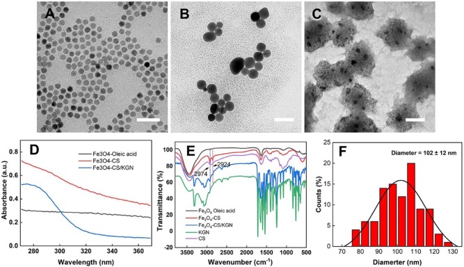
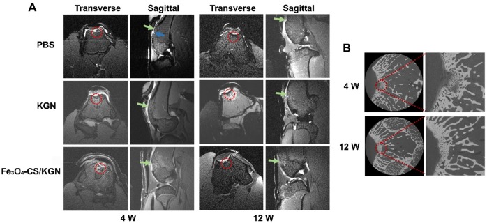
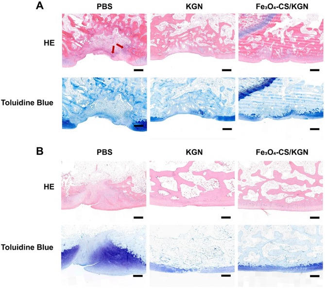
Chondral and osteochondral defects caused by trauma or pathological changes, commonly progress into total joint degradation, even resulting in disability. The cartilage restoration is a great challenge because of its avascularity and limited proliferative ability. Additionally, precise diagnosis using non-invasive detection techniques is challenging, which increases problems associated with chondral disease treatment. To achieve a theranostic goal, we used an integrated strategy that relies on exploiting a multifunctional nanoprobe based on chitosan-modified Fe3O4 nanoparticles, which spontaneously self-assemble with the oppositely charged small molecule growth factor, kartogenin (KGN). This nanoprobe was used to obtain distinctively brighter T-weighted magnetic resonance (MR) imaging, allowing its use as a positive contrast agent, and could be applied to obtain accurate diagnosis and osteochondral regeneration therapy. This nanoprobe was first investigated using adipose tissue-derived stem cells (ADSCs), and was found to be a novel positive contrast agent that also plays a significant role in stimulating ADSCs differentiation into chondrocytes. This self-assembled probe was not only biocompatible both and , contributing to cellular internalization, but was also used to successfully make distinction of normal/damaged tissue in T-weighted MR imaging. This novel combination was systematically shown to be biosafe via the decrement of apparent MR signals and elimination of ferroferric oxide over a 12-week regeneration period. Here, we established a novel method for osteochondral disease diagnosis and reconstruction. Using the FeO-CS/KGN nanoprobe, it is easy to distinguish the defect position, and it could act as a tool for dynamic observation as well as a stem cell-based therapy for directionally chondral differentiation.
由于创伤或病理变化导致的软骨和软骨下骨缺损,通常会导致全关节降解,甚至导致残疾。由于软骨的无血管性和有限的增殖能力,软骨修复是一个巨大的挑战。此外,使用非侵入性检测技术进行精确诊断具有挑战性,这增加了与软骨疾病治疗相关的问题。为了实现治疗诊断一体化的目标,我们采用了一种基于壳聚糖修饰的 Fe3O4 纳米粒子的多功能纳米探针的综合策略,该纳米粒子可自发与带相反电荷的小分子生长因子(KGN)自组装。该纳米探针用于获得明显更亮的 T 加权磁共振(MR)成像,可作为阳性对比剂使用,并可用于进行准确的诊断和软骨下再生治疗。首先在脂肪组织来源的干细胞(ADSCs)中研究了该纳米探针,发现其是一种新型的阳性对比剂,在刺激 ADSCs 向软骨细胞分化方面也起着重要作用。这种自组装探针不仅具有良好的生物相容性和细胞内化作用,还可用于 T 加权 MR 成像中正常/损伤组织的区分。通过在 12 周的再生期内降低明显的 MR 信号和消除四氧化三铁,该新型探针被系统地证明是生物安全的。在这里,我们建立了一种用于诊断和重建骨软骨疾病的新方法。使用 FeO-CS/KGN 纳米探针,很容易区分缺陷位置,并且可以作为动态观察的工具以及用于定向软骨分化的基于干细胞的治疗方法。