Liu Xincheng, Zhang Zhao, Shi Yubo, Meng Xingxing, Qiu Zhennan, Qu Xiaoli, Dang Jingyi, Zhang Yushen, Sun Liguo, Wang Lei, Zhu Dongze, Mi Zhenzhou, He Jiankang, Fan Hongbin
Department of Orthopedic Surgery, Xijing Hospital, the Fourth Military Medical University, Xi'an, China.
Department of Aerospace Hygiene, the Fourth Military Medical University, Xi'an, China.
Ann Transl Med. 2022 Jul;10(13):743. doi: 10.21037/atm-22-2796.
Osteoarthritis (OA) is a common degenerative disease. Chondrocyte dedifferentiation can accelerate the progress of OA. Three-dimensional printing (3DP) is widely used in tissue regeneration applications. A three-dimensional (3D) culture system with 3D printed scaffolds could reduce the dedifferentiation of chondrocytes during passages, which would be a potential method for chondrocyte expansion.
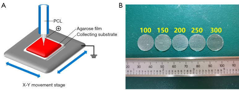
The viability and proliferation of chondrocytes on scaffolds and effects of scaffolds with 100, 150, 200, 250 or 300 µm spacing on chondrocyte dedifferentiation were analyzed . The morphology of scaffolds and cell/scaffold constructs was observed by scanning electron microscopy (SEM). Glycosaminoglycan (GAG) was evaluated by Alcian blue staining. The effects of different spacing on chondrocyte dedifferentiation were evaluated by the messenger RNA (mRNA) and protein levels of cartilage-related genes.
With more binding sites, the proliferation and viability of chondrocytes on scaffolds with 100 and 150 µm spacing were better than those with 200, 250 and 300 µm spacing on day 1, but this advantage diminished over time. The histology and quantitative real-time polymerase chain reaction (qRT-PCR) results showed that 200 µm spacing inhibits chondrocyte dedifferentiation better.
3D printed scaffolds with 200 µm spacing can inhibit chondrocyte dedifferentiation, providing a basis for the future study of 3D printed scaffolds as an effective method for chondrocyte expansion.
骨关节炎(OA)是一种常见的退行性疾病。软骨细胞去分化会加速OA的进展。三维打印(3DP)广泛应用于组织再生领域。具有三维打印支架的三维(3D)培养系统可减少传代过程中软骨细胞的去分化,这将是软骨细胞扩增的一种潜在方法。
分析软骨细胞在支架上的活力和增殖情况,以及间距为100、150、200、250或300 µm的支架对软骨细胞去分化的影响。通过扫描电子显微镜(SEM)观察支架及细胞/支架构建体的形态。用阿尔辛蓝染色评估糖胺聚糖(GAG)。通过软骨相关基因的信使核糖核酸(mRNA)和蛋白质水平评估不同间距对软骨细胞去分化的影响。
结合位点越多,间距为100和150 µm的支架上软骨细胞在第1天的增殖和活力优于间距为200、250和300 µm的支架,但这种优势会随时间减弱。组织学和定量实时聚合酶链反应(qRT-PCR)结果表明,200 µm的间距对软骨细胞去分化的抑制效果更好。
间距为200 µm的三维打印支架可抑制软骨细胞去分化,为未来将三维打印支架作为软骨细胞扩增的有效方法的研究提供了依据。