Wake Forest School of Medicine, Wake Forest Institute for Regenerative Medicine, Wake Forest Baptist Medical Center, Winston-Salem, North Carolina.
School of Medicine, Meharry Medical College, Nashville, Tennessee.
Biotechnol Bioeng. 2020 Aug;117(8):2516-2526. doi: 10.1002/bit.27379. Epub 2020 May 23.
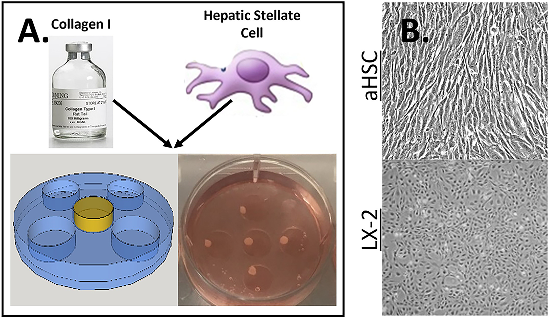
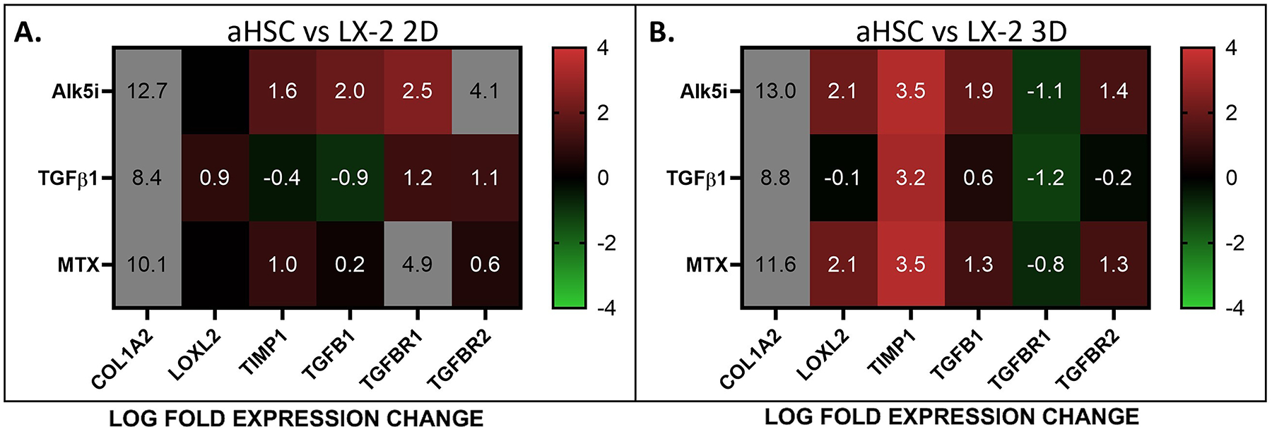
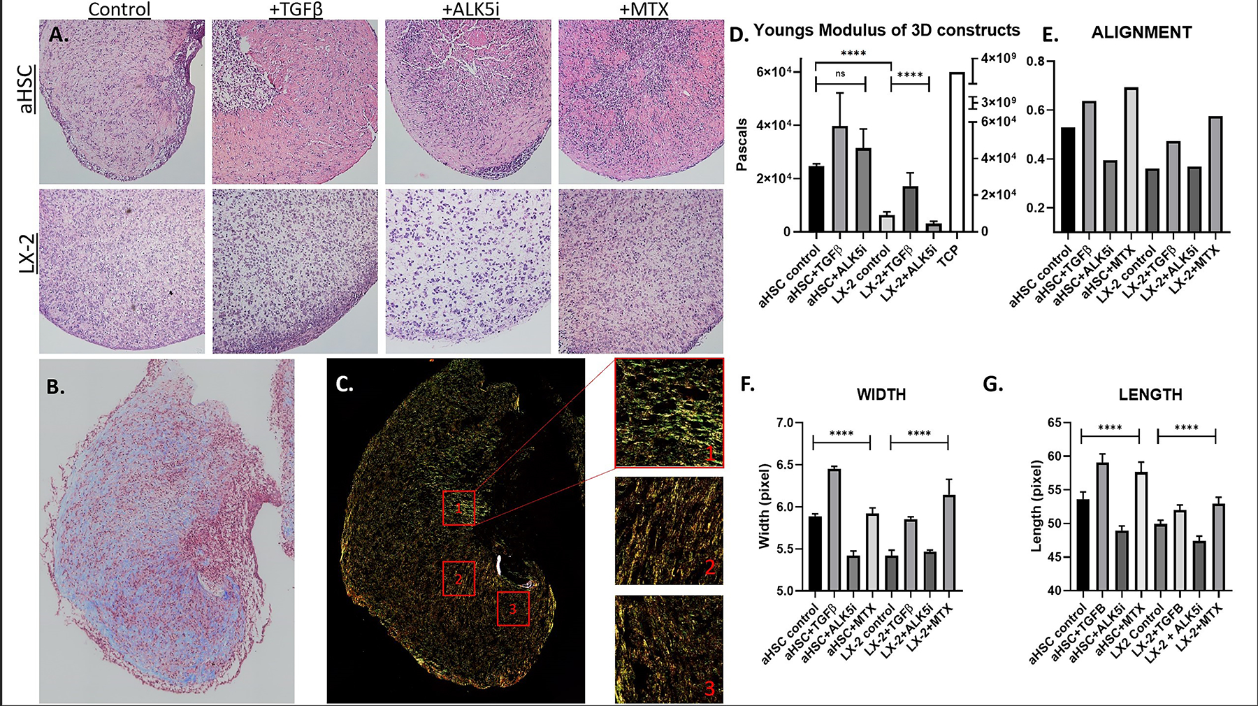
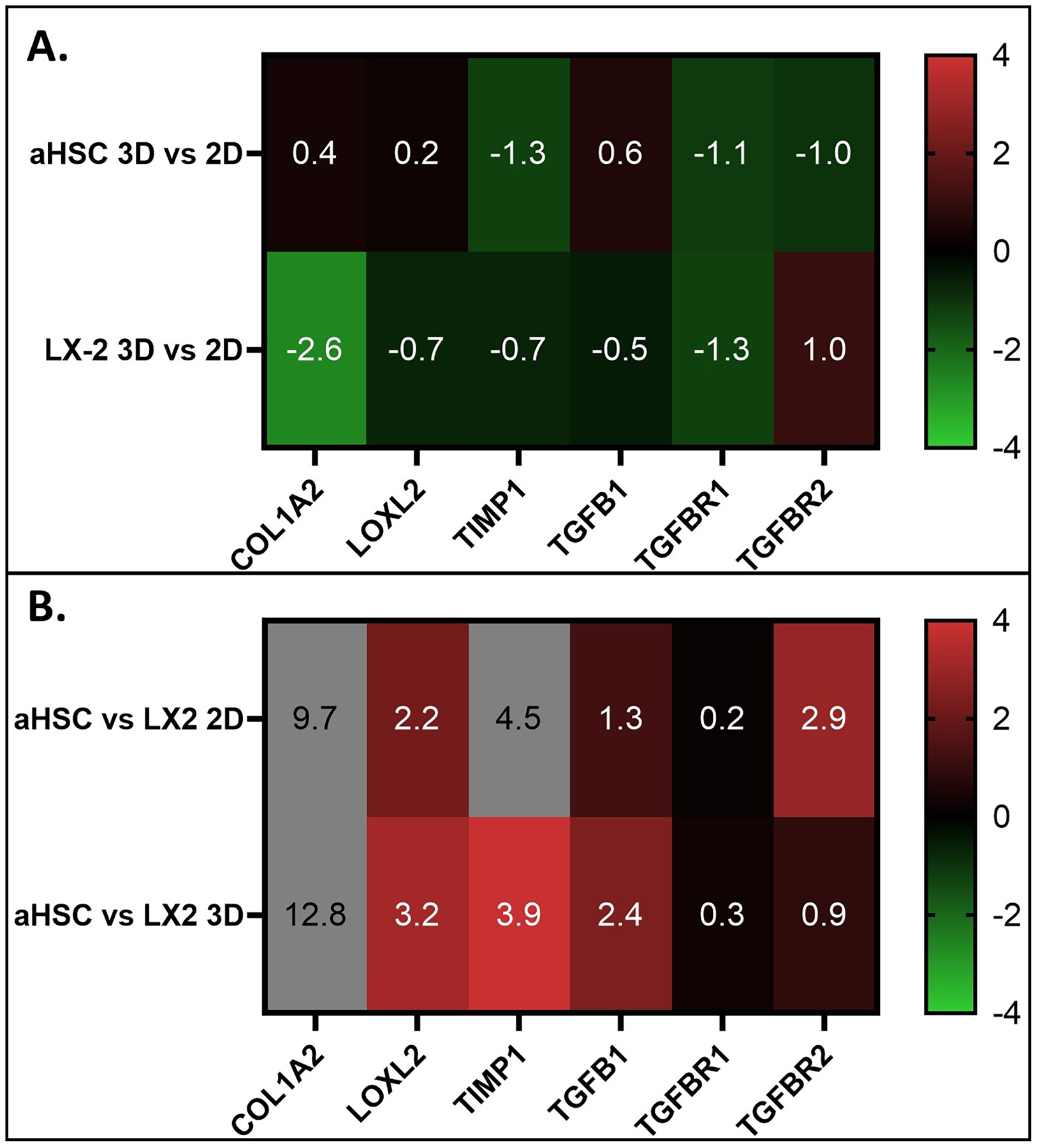
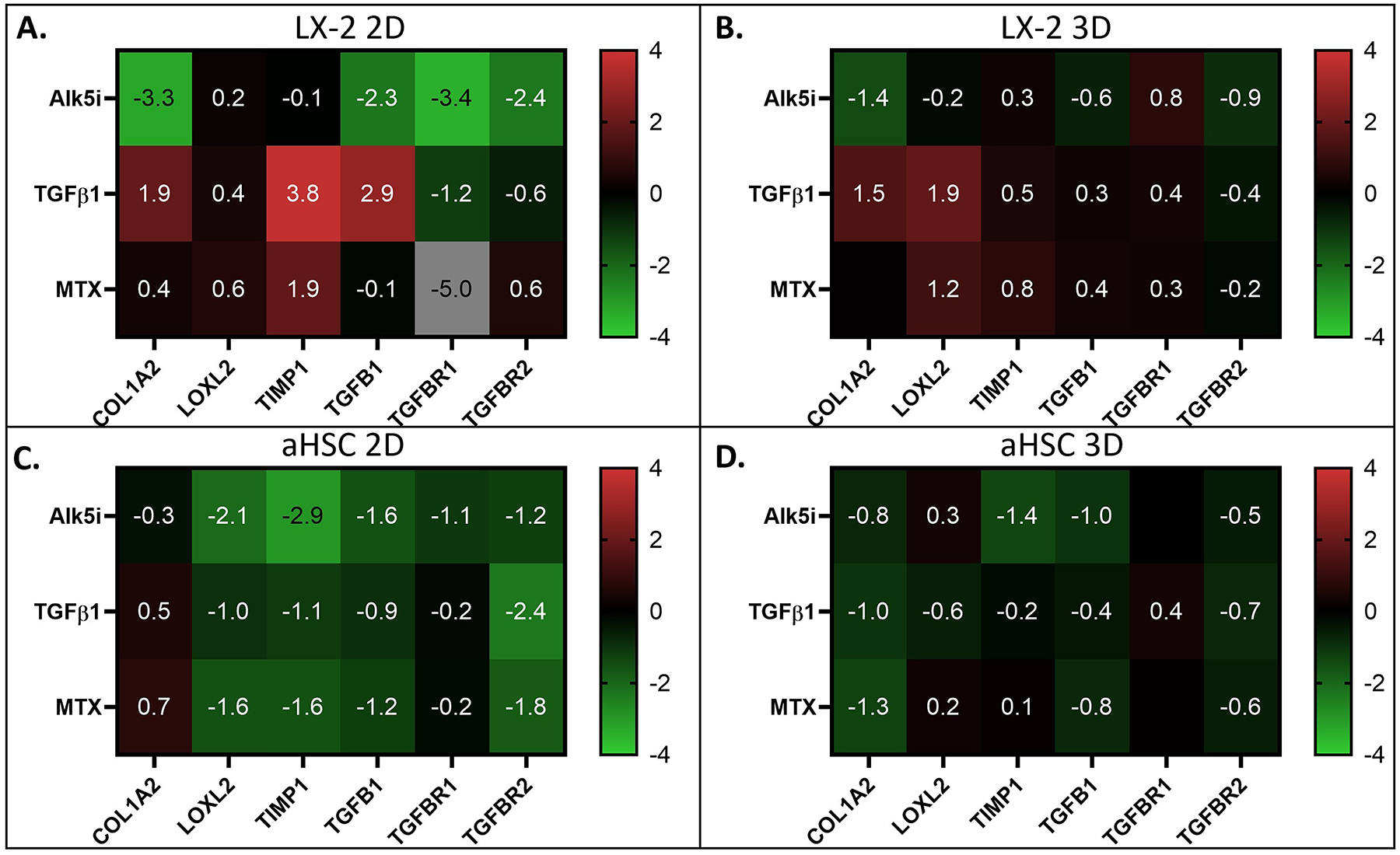
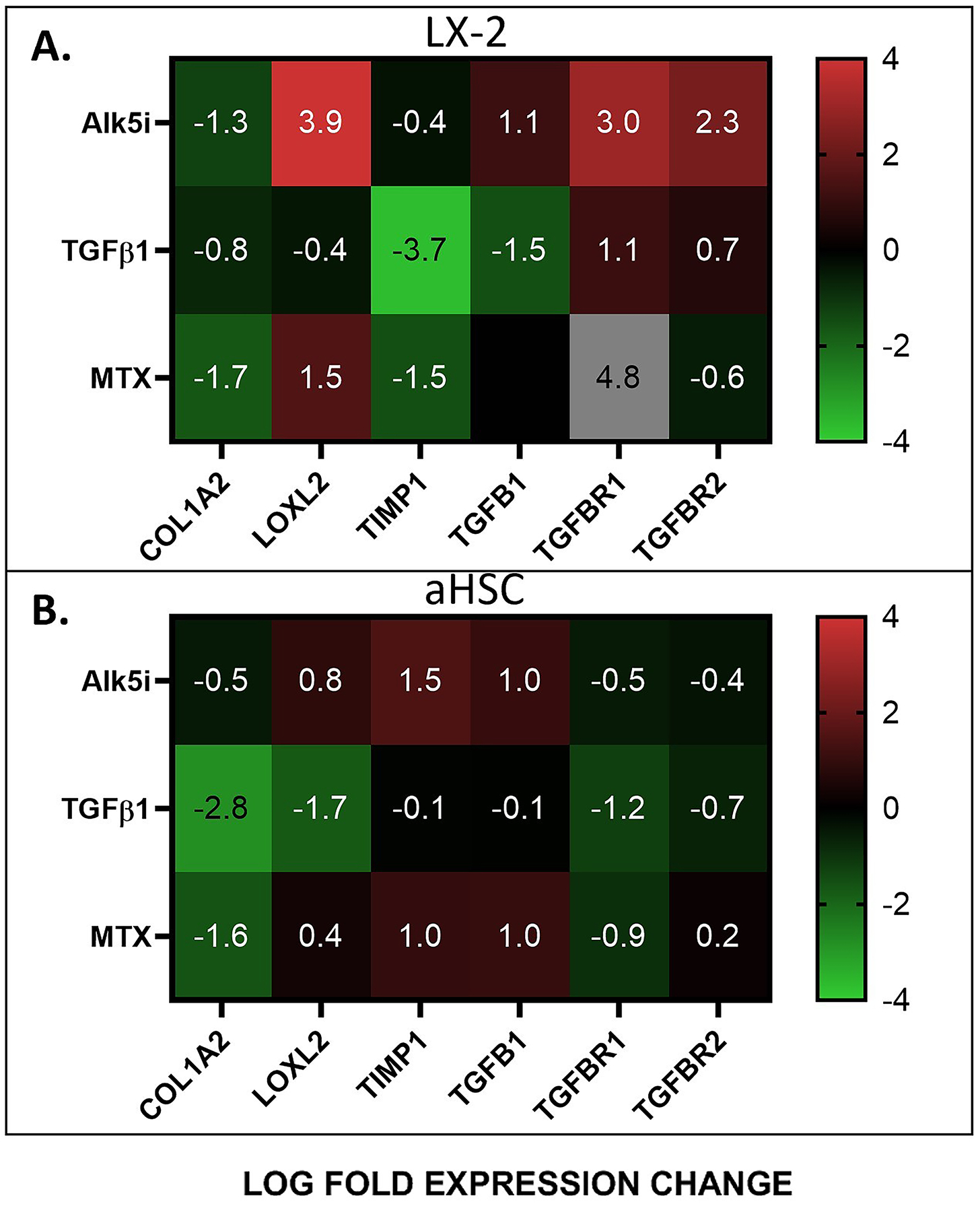
Liver fibrosis occurs in most cases of chronic liver disease, which are somewhat common, but also a potentially deadly group of diseases. In vitro modeling of liver fibrosis relies primarily on the isolation of in vivo activated hepatic stellate cells (aHSCs) and studying them in standard tissue culture dishes (two-dimensional [2D]). In contrast, modeling of fibrosis in a biofabricated three-dimensional (3D) construct allows us to study changes to the environment, such as extracellular matrix (ECM) composition and structure, and tissue rigidity. In the current study, we used aHSCs produced through subcultures in 2D and encapsulated them in a 3D collagen gel to form spherical constructs. In parallel, and as a comparison, we used an established HSC line, LX-2, representing early and less severe fibrosis. Compared with LX-2 cells, the aHSCs created a stiffer environment and expressed higher levels of TIMP1 and LOXL2, all of which are indicative of advanced liver fibrosis. Collectively, this study presents a fibrosis model that could be incorporated with multi-cellular models to more accurately reflect the effects of a severe fibrotic environment on liver function.
肝纤维化发生于大多数慢性肝病,这些疾病较为常见,但也是一组潜在致命的疾病。体外肝纤维化模型主要依赖于体内激活的肝星状细胞(aHSCs)的分离,并在标准组织培养皿(二维 [2D])中对其进行研究。相比之下,生物制造的三维(3D)构建体中的纤维化模型使我们能够研究环境的变化,如细胞外基质(ECM)的组成和结构以及组织硬度。在本研究中,我们使用通过 2D 传代培养产生的 aHSCs,并将其包裹在 3D 胶原凝胶中形成球形构建体。同时,作为比较,我们使用了一个已建立的 HSC 系 LX-2,代表早期和较轻的纤维化。与 LX-2 细胞相比,aHSCs 创造了一个更硬的环境,并表达了更高水平的 TIMP1 和 LOXL2,所有这些都表明存在晚期肝纤维化。总的来说,这项研究提出了一种纤维化模型,可以与多细胞模型结合使用,以更准确地反映严重纤维化环境对肝功能的影响。