• 文献检索
  • 文档翻译
  • 深度研究
  • 学术资讯
  • Suppr Zotero 插件Zotero 插件
  • 邀请有礼
  • 套餐&价格
  • 历史记录
应用&插件
Suppr Zotero 插件Zotero 插件浏览器插件Mac 客户端Windows 客户端微信小程序
定价
高级版会员购买积分包购买API积分包
服务
文献检索文档翻译深度研究API 文档MCP 服务
关于我们
关于 Suppr公司介绍联系我们用户协议隐私条款
关注我们

Suppr 超能文献

核心技术专利:CN118964589B侵权必究
粤ICP备2023148730 号-1Suppr @ 2026

文献检索

告别复杂PubMed语法,用中文像聊天一样搜索,搜遍4000万医学文献。AI智能推荐,让科研检索更轻松。

立即免费搜索

文件翻译

保留排版,准确专业,支持PDF/Word/PPT等文件格式,支持 12+语言互译。

免费翻译文档

深度研究

AI帮你快速写综述,25分钟生成高质量综述,智能提取关键信息,辅助科研写作。

立即免费体验

使用肝细胞、原代肝窦内皮细胞、肝星状细胞和巨噬细胞生成肝纤维化类器官。

Generation of Fibrotic Liver Organoids Using Hepatocytes, Primary Liver Sinusoidal Endothelial Cells, Hepatic Stellate Cells, and Macrophages.

机构信息

Regeneration Medicine Research Center, Yonsei University Wonju College of Medicine, Wonju 26426, Republic of Korea.

Department of Surgery, Yonsei University Wonju College of Medicine, Wonju 26426, Republic of Korea.

出版信息

Cells. 2023 Oct 24;12(21):2514. doi: 10.3390/cells12212514.

DOI:10.3390/cells12212514
PMID:37947592
原文链接:https://pmc.ncbi.nlm.nih.gov/articles/PMC10647544/
Abstract

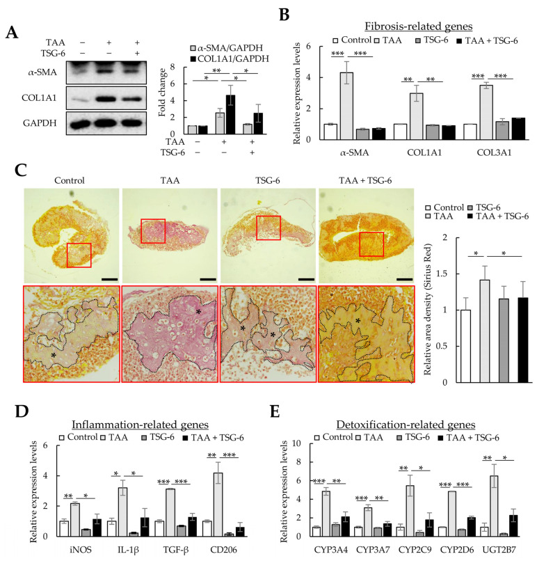
Liver organoids generated with single or multiple cell types have been used to investigate liver fibrosis development, toxicity, pathogenesis, and drug screening. However, organoid generation is limited by the availability of cells isolated from primary tissues or differentiated from various stem cells. To ensure cell availability for organoid formation, we investigated whether liver organoids could be generated with cell-line-based Huh-7 hepatocellular carcinoma cells, macrophages differentiated from THP-1 monocytes, and LX-2 hepatic stellate cells (HSCs) and primary liver sinusoidal endothelial cells (LSECs). In liver organoids, hepatocyte-, LSEC-, macrophage-, and HSC-related gene expression increased relative to that in two-dimensional (2D)-cultured Huh-7/LSEC/THP-1/LX-2 cells without Matrigel. Thioacetamide (TAA) increased α-smooth muscle actin expression in liver organoids but not in 2D-cultured cells, whereas in TAA-treated organoids, the expression of hepatic and LSEC markers decreased and that of macrophage and HSC markers increased. TAA-induced fibrosis was suppressed by treatment with N-acetyl-L-cysteine or tumor-necrosis-factor-stimulated gene 6 protein. The results showed that liver toxicants could induce fibrotic and inflammatory responses in liver organoids comprising Huh-7/LSEC/macrophages/LX-2 cells, resulting in fibrotic liver organoids. We propose that cell-line-based organoids can be used for disease modeling and drug screening to improve liver fibrosis treatment.

摘要

利用单一或多种细胞类型生成的肝类器官已被用于研究肝纤维化的发展、毒性、发病机制和药物筛选。然而,类器官的生成受到从原代组织中分离或从各种干细胞中分化得到的细胞的可用性的限制。为了确保类器官形成所需的细胞可用性,我们研究了是否可以使用基于细胞系的 Huh-7 肝癌细胞、从 THP-1 单核细胞分化而来的巨噬细胞以及 LX-2 肝星状细胞 (HSCs) 和原代肝窦内皮细胞 (LSECs) 生成肝类器官。在肝类器官中,与无 Matrigel 的二维 (2D) 培养 Huh-7/LSEC/THP-1/LX-2 细胞相比,肝细胞、LSEC、巨噬细胞和 HSC 相关基因的表达增加。硫代乙酰胺 (TAA) 增加了肝类器官中α-平滑肌肌动蛋白的表达,但在 2D 培养的细胞中没有增加,而在 TAA 处理的类器官中,肝和 LSEC 标志物的表达降低,巨噬细胞和 HSC 标志物的表达增加。N-乙酰-L-半胱氨酸或肿瘤坏死因子刺激基因 6 蛋白的治疗抑制了 TAA 诱导的纤维化。结果表明,肝毒物可诱导包含 Huh-7/LSEC/巨噬细胞/LX-2 细胞的肝类器官发生纤维性和炎症反应,导致纤维性肝类器官。我们提出,基于细胞系的类器官可用于疾病建模和药物筛选,以改善肝纤维化的治疗。

https://cdn.ncbi.nlm.nih.gov/pmc/blobs/9c88/10647544/ff7d0ebadfcd/cells-12-02514-g008.jpg
https://cdn.ncbi.nlm.nih.gov/pmc/blobs/9c88/10647544/1a8d830e712b/cells-12-02514-g001.jpg
https://cdn.ncbi.nlm.nih.gov/pmc/blobs/9c88/10647544/837da1d5177b/cells-12-02514-g002.jpg
https://cdn.ncbi.nlm.nih.gov/pmc/blobs/9c88/10647544/b4a265cd73f3/cells-12-02514-g003.jpg
https://cdn.ncbi.nlm.nih.gov/pmc/blobs/9c88/10647544/c14c91b017b9/cells-12-02514-g004.jpg
https://cdn.ncbi.nlm.nih.gov/pmc/blobs/9c88/10647544/68609ce6d1d5/cells-12-02514-g005.jpg
https://cdn.ncbi.nlm.nih.gov/pmc/blobs/9c88/10647544/99bd62d940b1/cells-12-02514-g006.jpg
https://cdn.ncbi.nlm.nih.gov/pmc/blobs/9c88/10647544/15675d179a9a/cells-12-02514-g007.jpg
https://cdn.ncbi.nlm.nih.gov/pmc/blobs/9c88/10647544/ff7d0ebadfcd/cells-12-02514-g008.jpg

相似文献

1
Generation of Fibrotic Liver Organoids Using Hepatocytes, Primary Liver Sinusoidal Endothelial Cells, Hepatic Stellate Cells, and Macrophages.使用肝细胞、原代肝窦内皮细胞、肝星状细胞和巨噬细胞生成肝纤维化类器官。
Cells. 2023 Oct 24;12(21):2514. doi: 10.3390/cells12212514.
2
FXR agonist obeticholic acid reduces hepatic inflammation and fibrosis in a rat model of toxic cirrhosis.法尼醇 X 受体激动剂奥贝胆酸可减少大鼠中毒性肝硬化模型中的肝脏炎症和纤维化。
Sci Rep. 2016 Sep 16;6:33453. doi: 10.1038/srep33453.
3
Cooperation of liver cells in health and disease.健康与疾病状态下肝细胞的协作。
Adv Anat Embryol Cell Biol. 2001;161:III-XIII, 1-151. doi: 10.1007/978-3-642-56553-3.
4
AMP-activated protein kinase activator, HL156A reduces thioacetamide-induced liver fibrosis in mice and inhibits the activation of cultured hepatic stellate cells and macrophages.AMP 激活的蛋白激酶激活剂 HL156A 可减轻硫代乙酰胺诱导的小鼠肝纤维化,并抑制培养的肝星状细胞和巨噬细胞的激活。
Int J Oncol. 2016 Oct;49(4):1407-14. doi: 10.3892/ijo.2016.3627. Epub 2016 Jul 21.
5
Tumor necrosis factor-inducible gene 6 reprograms hepatic stellate cells into stem-like cells, which ameliorates liver damage in mouse.肿瘤坏死因子诱导基因 6 可将肝星状细胞重编程为干细胞样细胞,从而改善小鼠的肝损伤。
Biomaterials. 2019 Oct;219:119375. doi: 10.1016/j.biomaterials.2019.119375. Epub 2019 Jul 22.
6
Hepatitis C virus core protein induces dysfunction of liver sinusoidal endothelial cell by down-regulation of silent information regulator 1.丙型肝炎病毒核心蛋白通过下调沉默信息调节因子 1 诱导肝窦内皮细胞功能障碍。
J Med Virol. 2018 May;90(5):926-935. doi: 10.1002/jmv.25034. Epub 2018 Feb 2.
7
Pro-fibrotic compounds induce stellate cell activation, ECM-remodelling and Nrf2 activation in a human 3D-multicellular model of liver fibrosis.促纤维化化合物在人肝纤维化三维多细胞模型中诱导星状细胞活化、细胞外基质重塑和Nrf2活化。
PLoS One. 2017 Jun 30;12(6):e0179995. doi: 10.1371/journal.pone.0179995. eCollection 2017.
8
Endothelial GATA4 controls liver fibrosis and regeneration by preventing a pathogenic switch in angiocrine signaling.内皮细胞 GATA4 通过防止血管生成信号的致病转换来控制肝纤维化和再生。
J Hepatol. 2021 Feb;74(2):380-393. doi: 10.1016/j.jhep.2020.08.033. Epub 2020 Sep 9.
9
Inhibition of protein arginine methyltransferase 1 alleviates liver fibrosis by attenuating the activation of hepatic stellate cells in mice.抑制蛋白质精氨酸甲基转移酶1可通过减弱小鼠肝星状细胞的激活来减轻肝纤维化。
FASEB J. 2022 Sep;36(9):e22489. doi: 10.1096/fj.202200238R.
10
Interferon-β Overexpression in Adipose Tissue-Derived Stem Cells Induces HepG2 and Macrophage Cell Death in Liver Tumor Organoids via Induction of TNF-Related Apoptosis-Inducing Ligand Expression.脂肪组织来源的干细胞中干扰素-β的过表达通过诱导肿瘤坏死因子相关凋亡诱导配体表达诱导肝癌类器官中的 HepG2 和巨噬细胞死亡。
Int J Mol Sci. 2024 Jan 22;25(2):1325. doi: 10.3390/ijms25021325.

引用本文的文献

1
Decoding the hepatic fibrosis-hepatocellular carcinoma axis: from mechanisms to therapeutic opportunities.解读肝纤维化-肝细胞癌轴:从机制到治疗机遇
Hepatol Int. 2025 Jul 1. doi: 10.1007/s12072-025-10838-y.
2
Recent Advances in Three-Dimensional In Vitro Models for Studies of Liver Fibrosis.用于肝纤维化研究的三维体外模型的最新进展
Tissue Eng Regen Med. 2025 May 13. doi: 10.1007/s13770-025-00719-8.
3
Pharmacotherapy of Liver Fibrosis and Hepatitis: Recent Advances.肝纤维化和肝炎的药物治疗:最新进展

本文引用的文献

1
Antifibrotic TSG-6 Expression Is Synergistically Increased in Both Cells during Coculture of Mesenchymal Stem Cells and Macrophages via the JAK/STAT Signaling Pathway.细胞间共培养通过 JAK/STAT 信号通路协同上调间充质干细胞和巨噬细胞中抗纤维化 TSG-6 的表达。
Int J Mol Sci. 2022 Oct 28;23(21):13122. doi: 10.3390/ijms232113122.
2
Liver Organoids: Recent Developments, Limitations and Potential.肝脏类器官:最新进展、局限性与潜力
Front Med (Lausanne). 2021 May 5;8:574047. doi: 10.3389/fmed.2021.574047. eCollection 2021.
3
Hepatic macrophages in liver homeostasis and diseases-diversity, plasticity and therapeutic opportunities.
Pharmaceuticals (Basel). 2024 Dec 20;17(12):1724. doi: 10.3390/ph17121724.
4
The use of organoids in creating immune microenvironments and treating gynecological tumors.类器官在创建免疫微环境和治疗妇科肿瘤中的应用。
J Transl Med. 2024 Sep 23;22(1):856. doi: 10.1186/s12967-024-05649-y.
5
Interferon-β Overexpression in Adipose Tissue-Derived Stem Cells Induces HepG2 and Macrophage Cell Death in Liver Tumor Organoids via Induction of TNF-Related Apoptosis-Inducing Ligand Expression.脂肪组织来源的干细胞中干扰素-β的过表达通过诱导肿瘤坏死因子相关凋亡诱导配体表达诱导肝癌类器官中的 HepG2 和巨噬细胞死亡。
Int J Mol Sci. 2024 Jan 22;25(2):1325. doi: 10.3390/ijms25021325.
肝脏稳态中的肝巨噬细胞:多样性、可塑性和治疗机会。
Cell Mol Immunol. 2021 Jan;18(1):45-56. doi: 10.1038/s41423-020-00558-8. Epub 2020 Oct 12.
4
The Role of Sinusoidal Endothelial Cells in the Axis of Inflammation and Cancer Within the Liver.肝窦内皮细胞在肝脏炎症与癌症轴中的作用
Front Physiol. 2020 Aug 28;11:990. doi: 10.3389/fphys.2020.00990. eCollection 2020.
5
Liver Fibrosis: Mechanistic Concepts and Therapeutic Perspectives.肝纤维化:机制概念与治疗视角。
Cells. 2020 Apr 3;9(4):875. doi: 10.3390/cells9040875.
6
Liver-targeted delivery of TSG-6 by calcium phosphate nanoparticles for the management of liver fibrosis.通过磷酸钙纳米粒实现 TSG-6 的肝脏靶向递送,用于肝纤维化的治疗。
Theranostics. 2020 Jan 1;10(1):36-49. doi: 10.7150/thno.37301. eCollection 2020.
7
Human ESC-derived expandable hepatic organoids enable therapeutic liver repopulation and pathophysiological modeling of alcoholic liver injury.人胚胎干细胞衍生的可扩增肝类器官可实现治疗性肝再生以及酒精性肝损伤的病理生理建模。
Cell Res. 2019 Dec;29(12):1009-1026. doi: 10.1038/s41422-019-0242-8. Epub 2019 Oct 18.
8
Stellate Cells, Hepatocytes, and Endothelial Cells Imprint the Kupffer Cell Identity on Monocytes Colonizing the Liver Macrophage Niche.星状细胞、肝细胞和内皮细胞在单核细胞定殖肝脏巨噬细胞龛时赋予其库普弗细胞特征。
Immunity. 2019 Oct 15;51(4):638-654.e9. doi: 10.1016/j.immuni.2019.08.017. Epub 2019 Sep 24.
9
The cell biology of the hepatocyte: A membrane trafficking machine.肝细胞的细胞生物学:一种膜运输机器。
J Cell Biol. 2019 Jul 1;218(7):2096-2112. doi: 10.1083/jcb.201903090. Epub 2019 Jun 14.
10
Modeling Steatohepatitis in Humans with Pluripotent Stem Cell-Derived Organoids.利用多能干细胞衍生类器官模拟肝脂肪变性。
Cell Metab. 2019 Aug 6;30(2):374-384.e6. doi: 10.1016/j.cmet.2019.05.007. Epub 2019 May 30.