Department of Mathematical Sciences, National Chengchi University.
Department of Clinical Laboratory Sciences and Medical Biotechnology, National Taiwan University College of Medicine, Taipei, Taiwan.
PLoS Comput Biol. 2020 May 13;16(5):e1007883. doi: 10.1371/journal.pcbi.1007883. eCollection 2020 May.
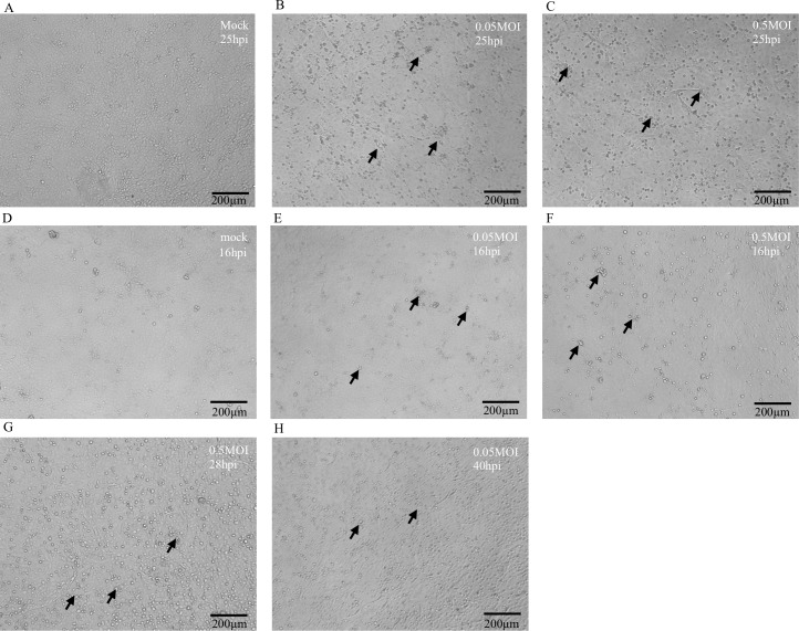
Cell culture remains as the golden standard for primary isolation of viruses in clinical specimens. In the current practice, researchers have to recognize the cytopathic effects (CPE) induced by virus infection and subsequently use virus-specific monoclonal antibody to confirm the presence of virus. Considering the broad applications of neural network in various fields, we aimed to utilize convolutional neural networks (CNN) to shorten the timing required for CPE identification and to improve the assay sensitivity. Based on the characteristics of influenza-induced CPE, a CNN model with larger sizes of filters and max-pooling kernels was constructed in the absence of transfer learning. A total of 601 images from mock-infected and influenza-infected MDCK cells were used to train the model. The performance of the model was tested by using extra 400 images and the percentage of correct recognition was 99.75%. To further examine the limit of our model in evaluating the changes of CPE overtime, additional 1190 images from a new experiment were used and the recognition rates at 16 hour (hr), 28 hr, and 40 hr post virus infection were 71.80%, 98.25%, and 87.46%, respectively. The specificity of our model, examined by images of MDCK cells infected by six other non-influenza viruses, was 100%. Hence, a simple CNN model was established to enhance the identification of influenza virus in clinical practice.
细胞培养仍然是临床标本中病毒初次分离的金标准。在目前的实践中,研究人员必须识别病毒感染引起的细胞病变效应(CPE),然后使用病毒特异性单克隆抗体来确认病毒的存在。鉴于神经网络在各个领域的广泛应用,我们旨在利用卷积神经网络(CNN)来缩短识别 CPE 所需的时间,并提高检测的灵敏度。基于流感引起的 CPE 的特征,在没有迁移学习的情况下构建了一个具有更大滤波器和最大池化核的 CNN 模型。该模型使用 601 张来自mock 感染和流感感染的 MDCK 细胞的图像进行训练。使用另外 400 张图像来测试模型的性能,正确识别的百分比为 99.75%。为了进一步检查我们的模型在评估 CPE 随时间变化的极限,使用来自新实验的另外 1190 张图像,在感染病毒后 16 小时(hr)、28 小时和 40 小时的识别率分别为 71.80%、98.25%和 87.46%。我们的模型的特异性,通过感染六种其他非流感病毒的 MDCK 细胞的图像进行检查,为 100%。因此,建立了一个简单的 CNN 模型来增强临床实践中流感病毒的识别。