HemanthaKumar G, Sathish Muthu
Department of Orthopaedics and Traumatology, Rajiv Gandhi Government General Hospital, Chennai, Tamil Nadu, India.
J Orthop Case Rep. 2019;9(4):101-105. doi: 10.13107/jocr.2019.v09.i04.1500.
Chondromyxoid fibroma (CMF) is a benign rare bone tumor of slow-growing nature arising from chondroblastic derivation. CMF in most of the cases is a diagnosis of exclusion, and in this case report, we differentiate the histological and radiological findings of CMF and difficulties in diagnosis of CMF from potential differential diagnosis.
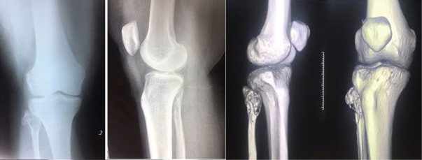
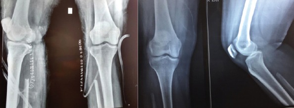
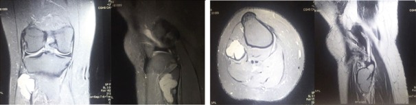
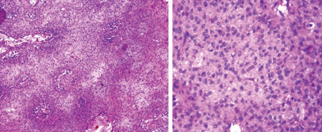
A 38-year-old female patient presented with a history of limping for 5 months and on evaluation revealed an expansile osteolytic lesion in fibular head with septations and soft tissue component. Excision biopsy was done. Histological examination revealed a cellular neoplasm arranged as vague nodules in chondroid background with occasional mitotic figures and giant cells in periphery without any calcification. To rule out chondroblastoma, S-100 and epithelial markers were done which was negative establishing diagnosis of CMF by exclusion.
CMF is often misdiagnosed being a radiological and pathological mimicker. Histology remains key to diagnosis. En bloc resection remains the mainstay of management in expendable bone-like fibula.
软骨黏液样纤维瘤(CMF)是一种起源于软骨母细胞的良性罕见骨肿瘤,生长缓慢。大多数情况下,CMF是一种排除性诊断,在本病例报告中,我们区分了CMF的组织学和放射学表现以及CMF诊断中的困难与潜在的鉴别诊断。
一名38岁女性患者有5个月跛行病史,评估发现腓骨头有一个膨胀性溶骨性病变,伴有分隔和软组织成分。进行了切除活检。组织学检查显示,肿瘤细胞呈细胞性,在软骨样背景中呈模糊结节状排列,偶见有丝分裂象,周边有巨细胞,无任何钙化。为排除软骨母细胞瘤,进行了S-100和上皮标志物检查,结果为阴性,通过排除法确诊为CMF。
CMF常因影像学和病理学表现相似而被误诊。组织学仍是诊断的关键。整块切除仍是像腓骨这样可切除骨的主要治疗方法。