• 文献检索
  • 文档翻译
  • 深度研究
  • 学术资讯
  • Suppr Zotero 插件Zotero 插件
  • 邀请有礼
  • 套餐&价格
  • 历史记录
应用&插件
Suppr Zotero 插件Zotero 插件浏览器插件Mac 客户端Windows 客户端微信小程序
定价
高级版会员购买积分包购买API积分包
服务
文献检索文档翻译深度研究API 文档MCP 服务
关于我们
关于 Suppr公司介绍联系我们用户协议隐私条款
关注我们

Suppr 超能文献

核心技术专利:CN118964589B侵权必究
粤ICP备2023148730 号-1Suppr @ 2026

文献检索

告别复杂PubMed语法,用中文像聊天一样搜索,搜遍4000万医学文献。AI智能推荐,让科研检索更轻松。

立即免费搜索

文件翻译

保留排版,准确专业,支持PDF/Word/PPT等文件格式,支持 12+语言互译。

免费翻译文档

深度研究

AI帮你快速写综述,25分钟生成高质量综述,智能提取关键信息,辅助科研写作。

立即免费体验

自发性蛛网膜下腔出血相关脑损伤的扩散张量成像研究:一项小型综述。

Diffusion Tensor Imaging Studies on Spontaneous Subarachnoid Hemorrhage-Related Brain Injury: A Mini-Review.

作者信息

Cho Min Kyeong, Jang Sung Ho

机构信息

Department of Physical Medicine and Rehabilitation, College of Medicine, Yeungnam University, Daegu, South Korea.

出版信息

Front Neurol. 2020 Apr 28;11:283. doi: 10.3389/fneur.2020.00283. eCollection 2020.

DOI:10.3389/fneur.2020.00283
PMID:32411076
原文链接:https://pmc.ncbi.nlm.nih.gov/articles/PMC7198780/
Abstract

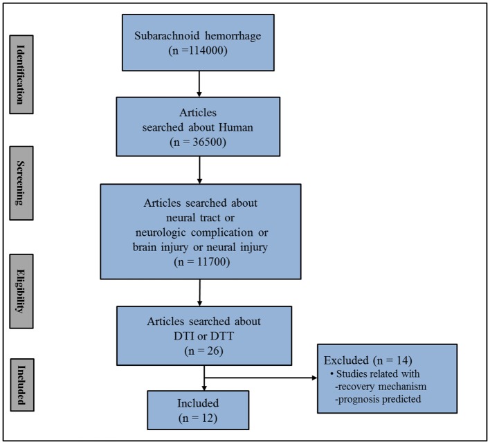
Accurate diagnosis of the presence and severity of neural injury in patients with subarachnoid hemorrhage (SAH) is important in neurorehabilitation because it is essential for establishing appropriate therapeutic strategies and developing a prognosis. Diffusion tensor imaging has a unique advantage in the identification of microstructural white matter abnormalities which are not usually detectable on conventional brain magnetic resonance imaging. In this mini-review article, 12 diffusion tensor imaging studies on SAH-related brain injury were reviewed. These studies have demonstrated SAH-related brain injuries in various neural tracts or structures including the cingulum, fornix, hippocampus, dorsolateral prefrontal region, corticospinal tract, mamillothalamic tract, corticoreticular pathway, ascending reticular activating system, Papez circuit, optic radiation, and subcortical white matter. We believe that these reviewed studies provide information that would be helpful in science-based neurorehabilitation of patients with SAH. Furthermore, the results of these reviewed studies would also be useful for clarification of the pathophysiological mechanisms associated with SAH-related brain injury. However, considering the large number of neural tracts or neural structures in the brain, more research on SAH-related brain injury in other neural tracts or structures should be encouraged.

摘要

准确诊断蛛网膜下腔出血(SAH)患者神经损伤的存在及严重程度在神经康复中至关重要,因为这对于制定合适的治疗策略和预测预后必不可少。扩散张量成像在识别微观结构白质异常方面具有独特优势,而这些异常在传统脑磁共振成像中通常无法检测到。在这篇小型综述文章中,对12项关于SAH相关脑损伤的扩散张量成像研究进行了综述。这些研究已证实在包括扣带束、穹窿、海马、背外侧前额叶区域、皮质脊髓束、乳头丘脑束、皮质网状通路、上行网状激活系统、帕佩兹环路、视辐射和皮质下白质等各种神经束或结构中存在SAH相关脑损伤。我们认为,这些综述研究提供的信息将有助于对SAH患者进行基于科学的神经康复。此外,这些综述研究的结果对于阐明与SAH相关脑损伤相关的病理生理机制也将是有用的。然而,考虑到大脑中神经束或神经结构的数量众多,应鼓励对其他神经束或结构中SAH相关脑损伤进行更多研究。

https://cdn.ncbi.nlm.nih.gov/pmc/blobs/54c6/7198780/de4245e9a1ad/fneur-11-00283-g0002.jpg
https://cdn.ncbi.nlm.nih.gov/pmc/blobs/54c6/7198780/49e08bae024b/fneur-11-00283-g0001.jpg
https://cdn.ncbi.nlm.nih.gov/pmc/blobs/54c6/7198780/de4245e9a1ad/fneur-11-00283-g0002.jpg
https://cdn.ncbi.nlm.nih.gov/pmc/blobs/54c6/7198780/49e08bae024b/fneur-11-00283-g0001.jpg
https://cdn.ncbi.nlm.nih.gov/pmc/blobs/54c6/7198780/de4245e9a1ad/fneur-11-00283-g0002.jpg

相似文献

1
Diffusion Tensor Imaging Studies on Spontaneous Subarachnoid Hemorrhage-Related Brain Injury: A Mini-Review.自发性蛛网膜下腔出血相关脑损伤的扩散张量成像研究:一项小型综述。
Front Neurol. 2020 Apr 28;11:283. doi: 10.3389/fneur.2020.00283. eCollection 2020.
2
A Review of Studies on the Role of Diffusion Tensor Magnetic Resonance Imaging Tractography in the Evaluation of the Fronto-Subcortical Circuit in Patients with Akinetic Mutism.磁共振弥散张量成像纤维束示踪技术在评估无动性缄默症患者额皮质下环路中的作用的研究综述
Med Sci Monit. 2022 Feb 19;28:e936251. doi: 10.12659/MSM.936251.
3
Diffusion tensor tractography characteristics of axonal injury in concussion/mild traumatic brain injury.脑震荡/轻度创伤性脑损伤中轴突损伤的扩散张量纤维束成像特征
Neural Regen Res. 2022 May;17(5):978-982. doi: 10.4103/1673-5374.324825.
4
Aneurysmal subarachnoid hemorrhage causes injury of the ascending reticular activating system: relation to consciousness.动脉瘤性蛛网膜下腔出血导致上行网状激活系统损伤:与意识的关系。
AJNR Am J Neuroradiol. 2015 Apr;36(4):667-71. doi: 10.3174/ajnr.A4203. Epub 2015 Jan 8.
5
Neural Tract Injuries Revealed by Diffusion Tensor Tractography in a Patient With Severe Heat Stroke.热射病患者的弥散张量纤维束成像揭示的神经束损伤。
Am J Phys Med Rehabil. 2020 Aug;99(8):e97-e100. doi: 10.1097/PHM.0000000000001291.
6
Demystifying White Matter Injury in the Unconscious Patients with Diffusion Tensor Imaging.利用扩散张量成像解读昏迷患者的白质损伤
Asian J Neurosurg. 2020 May 29;15(2):370-376. doi: 10.4103/ajns.AJNS_55_20. eCollection 2020 Apr-Jun.
7
Diffusion Tensor Tractography Studies on Injured Anterior Cingulum Recovery Mechanisms: A Mini-Review.扩散张量纤维束成像对前扣带回损伤恢复机制的研究:一项小型综述
Front Neurol. 2018 Dec 7;9:1073. doi: 10.3389/fneur.2018.01073. eCollection 2018.
8
Injuries of neural tracts in a patient with CADASIL: a diffusion tensor imaging study.伴有大脑常染色体显性动脉病伴皮质下梗死和白质脑病(CADASIL)患者的神经束损伤:一项扩散张量成像研究
BMC Neurol. 2015 Sep 28;15:176. doi: 10.1186/s12883-015-0434-x.
9
[Magnetic resonance diffusion tractography in the brain--its application and limitation].[大脑中的磁共振扩散张量成像——其应用与局限性]
Brain Nerve. 2007 May;59(5):467-76.
10
Association between scrub typhus encephalitis and diffusion tensor tractography detection of Papez circuit injury: A case report.恙虫病性脑炎与Papez环路损伤的弥散张量纤维束成像检测之间的关联:一例报告
World J Clin Cases. 2021 May 6;9(13):3194-3199. doi: 10.12998/wjcc.v9.i13.3194.

引用本文的文献

1
Characteristic parameter analysis of magnetic resonance diffusion tensor imaging in children with autism spectrum disorder: a retrospective study.孤独症谱系障碍儿童磁共振扩散张量成像特征参数分析:一项回顾性研究
BMC Pediatr. 2025 Jun 7;25(1):464. doi: 10.1186/s12887-025-05771-w.
2
White matter abnormalities in aneurysmal and angiographically negative subarachnoid hemorrhage: A diffusion kurtosis imaging study.颅内动脉瘤性和血管造影阴性蛛网膜下腔出血的脑白质异常:一项扩散峰度成像研究。
Neuroimage Clin. 2024;43:103662. doi: 10.1016/j.nicl.2024.103662. Epub 2024 Aug 30.
3
Non-Aneurysmal Perimesencephalic Subarachnoid Hemorrhage: A Literature Review.

本文引用的文献

1
Diagnostic Approach to Traumatic Axonal Injury of the Spinothalamic Tract in Individual Patients with Mild Traumatic Brain Injury.轻度创伤性脑损伤个体患者中脊髓丘脑束创伤性轴索损伤的诊断方法
Diagnostics (Basel). 2019 Nov 21;9(4):199. doi: 10.3390/diagnostics9040199.
2
The value of diffusion tensor imaging for differentiating autism spectrum disorder with language delay from developmental language disorder among toddlers.扩散张量成像在区分幼儿期伴有语言发育迟缓的自闭症谱系障碍与发育性语言障碍中的价值。
Medicine (Baltimore). 2019 Apr;98(14):e15058. doi: 10.1097/MD.0000000000015058.
3
Recovery process of bilaterally injured corticoreticulospinal tracts in a patient with subarachnoid hemorrhage: Case report.
非动脉瘤性中脑周围蛛网膜下腔出血:文献综述
Diagnostics (Basel). 2023 Mar 22;13(6):1195. doi: 10.3390/diagnostics13061195.
4
Altered hippocampal functional connectivity after the rupture of anterior communicating artery aneurysm.前交通动脉动脉瘤破裂后海马功能连接的改变。
Front Aging Neurosci. 2022 Nov 7;14:997231. doi: 10.3389/fnagi.2022.997231. eCollection 2022.
5
Advanced Imaging Techniques for Newly Diagnosed and Recurrent Gliomas.用于新诊断和复发性胶质瘤的先进成像技术
Front Neurosci. 2022 Feb 23;16:787755. doi: 10.3389/fnins.2022.787755. eCollection 2022.
6
Secondary White Matter Injury and Therapeutic Targets After Subarachnoid Hemorrhage.蛛网膜下腔出血后的继发性白质损伤及治疗靶点
Front Neurol. 2021 Jul 15;12:659740. doi: 10.3389/fneur.2021.659740. eCollection 2021.
7
Sleep deprivation aggravates brain injury after experimental subarachnoid hemorrhage via TLR4-MyD88 pathway.睡眠剥夺通过 TLR4-MyD88 通路加重实验性蛛网膜下腔出血后的脑损伤。
Aging (Albany NY). 2021 Jan 21;13(2):3101-3111. doi: 10.18632/aging.202503.
8
Understanding Cognitive Deficit After Subarachnoid Hemorrhage: A Memory Focused Approach.理解蛛网膜下腔出血后的认知缺陷:一种以记忆为重点的方法。
Cureus. 2020 Nov 16;12(11):e11513. doi: 10.7759/cureus.11513.
蛛网膜下腔出血患者双侧受损皮质网状脊髓束的恢复过程:病例报告
Medicine (Baltimore). 2018 Dec;97(50):e13401. doi: 10.1097/MD.0000000000013401.
4
Characterization of normal-appearing white matter in multiple sclerosis using quantitative susceptibility mapping in conjunction with diffusion tensor imaging.利用定量磁化率成像结合扩散张量成像对多发性硬化症中外观正常的白质进行特征描述。
Neuroradiology. 2019 Jan;61(1):71-79. doi: 10.1007/s00234-018-2137-7. Epub 2018 Dec 12.
5
Microstructural White Matter Abnormalities and Cognitive Impairment After Aneurysmal Subarachnoid Hemorrhage.脑动脉瘤性蛛网膜下腔出血后的脑微观结构白质异常与认知障碍。
Stroke. 2018 Sep;49(9):2040-2045. doi: 10.1161/STROKEAHA.118.021622.
6
Prediction of clinical outcome in subacute subarachnoid hemorrhage using diffusion tensor imaging.使用扩散张量成像预测亚急性蛛网膜下腔出血的临床结局
J Neurosurg. 2018 Apr 13;130(2):550-558. doi: 10.3171/2017.10.JNS171793. Print 2019 Feb 1.
7
Optic radiation injury in patients with aneurismal subarachnoid hemorrhage: A preliminary diffusion tensor imaging report.动脉瘤性蛛网膜下腔出血患者的视辐射损伤:一项初步的扩散张量成像报告。
Neural Regen Res. 2018 Mar;13(3):563-566. doi: 10.4103/1673-5374.228763.
8
Significant Injury of the Mammillothalamic Tract without Injury of the Corticospinal Tract After Aneurysmal Subarachnoid Hemorrhage: A Retrospective Diffusion Tensor Imaging Study.动脉瘤性蛛网膜下腔出血后乳头丘脑束严重损伤而皮质脊髓束未损伤:一项回顾性扩散张量成像研究
World Neurosurg. 2018 Jun;114:e624-e630. doi: 10.1016/j.wneu.2018.03.042. Epub 2018 Mar 13.
9
Injury of the Precommissural Fornix in a Patient with Subarachnoid Hemorrhage: A Case Report.一例蛛网膜下腔出血患者的穹窿连合前穹窿损伤:病例报告
J Stroke Cerebrovasc Dis. 2018 Jun;27(6):e98-e101. doi: 10.1016/j.jstrokecerebrovasdis.2017.12.045.
10
Diffusion Tensor Imaging (DTI).扩散张量成像(DTI)
Methods Mol Biol. 2018;1718:103-116. doi: 10.1007/978-1-4939-7531-0_7.