Department of Radiology and Biomedical Imaging, Yale School of Medicine, 300 Cedar Street, New Haven, CT, 06520, USA.
Institute of Radiology, Charité - Universitätsmedizin Berlin, corporate member of Freie Universität Berlin, Humboldt-Universität, and Berlin Institute of Health, 10117, Berlin, Germany.
Eur Radiol. 2020 Oct;30(10):5663-5673. doi: 10.1007/s00330-020-06931-5. Epub 2020 May 19.
To investigate the predictive value of quantifiable imaging and inflammatory biomarkers in patients with hepatocellular carcinoma (HCC) for the clinical outcome after drug-eluting bead transarterial chemoembolization (DEB-TACE) measured as volumetric tumor response and progression-free survival (PFS).
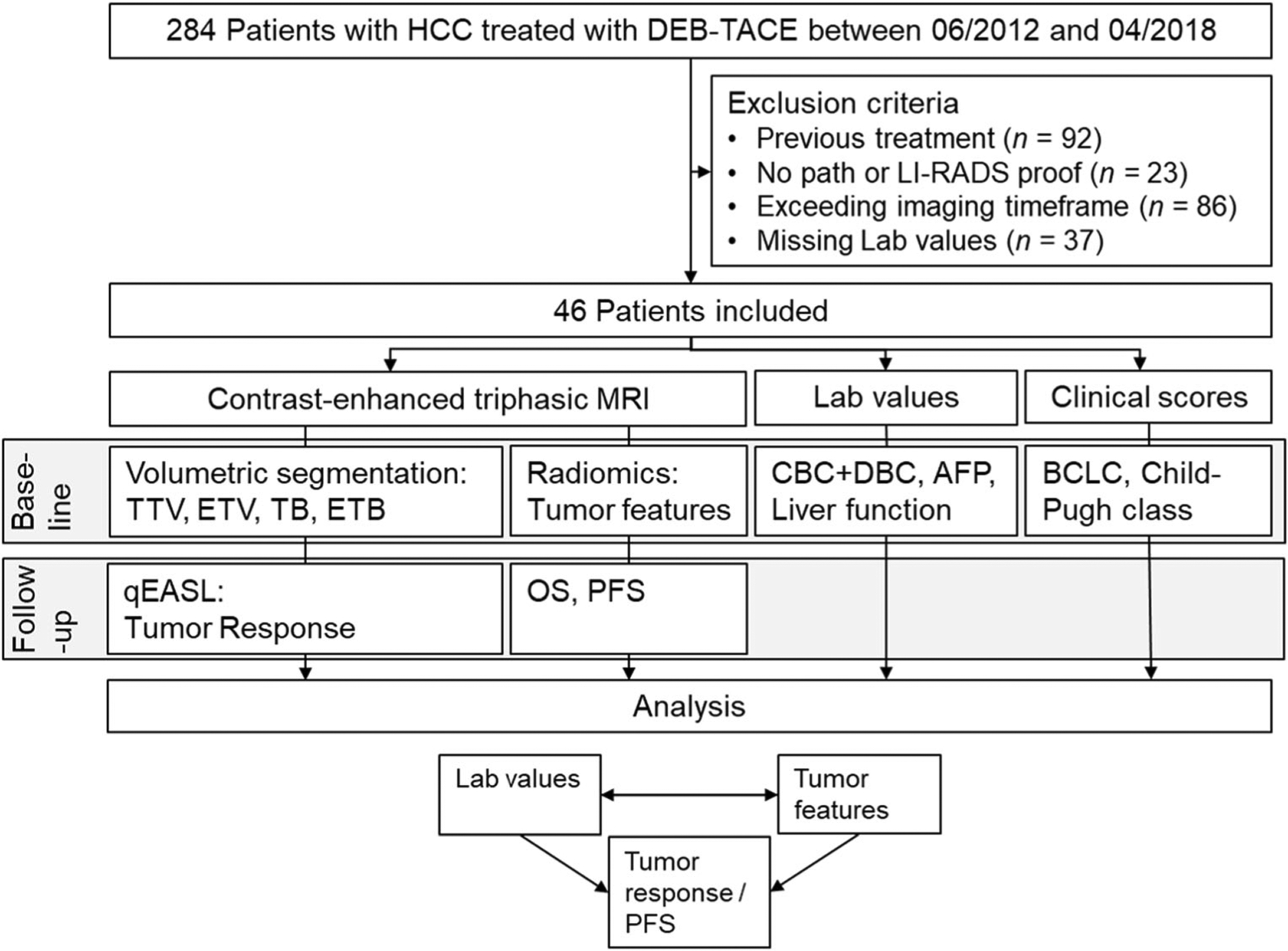
This retrospective study included 46 patients with treatment-naïve HCC who received DEB-TACE. Laboratory work-up prior to treatment included complete and differential blood count, liver function, and alpha-fetoprotein levels. Neutrophil-to-lymphocyte ratio (NLR) and platelet-to-lymphocyte ratio (PLR) were correlated with radiomic features extracted from pretreatment contrast-enhanced magnetic resonance imaging (MRI) and with tumor response according to quantitative European Association for the Study of the Liver (qEASL) criteria and progression-free survival (PFS) after DEB-TACE. Radiomic features included single nodular tumor growth measured as sphericity, dynamic contrast uptake behavior, arterial hyperenhancement, and homogeneity of contrast uptake. Statistics included univariate and multivariate linear regression, Cox regression, and Kaplan-Meier analysis.
Accounting for laboratory and clinical parameters, high baseline NLR and PLR were predictive of poorer tumor response (p = 0.014 and p = 0.004) and shorter PFS (p = 0.002 and p < 0.001). When compared to baseline imaging, high NLR and PLR correlated with non-spherical tumor growth (p = 0.001 and p < 0.001).
This study establishes the prognostic value of quantitative inflammatory biomarkers associated with aggressive non-spherical tumor growth and predictive of poorer tumor response and shorter PFS after DEB-TACE.
• In treatment-naïve hepatocellular carcinoma (HCC), high baseline platelet-to-lymphocyte ratio (PLR) and neutrophil-to-lymphocyte ratio (NLR) are associated with non-nodular tumor growth measured as low tumor sphericity. • High PLR and NLR are predictive of poorer volumetric enhancement-based tumor response and PFS after DEB-TACE in HCC. • This set of readily available, quantitative immunologic biomarkers can easily be implemented in clinical guidelines providing a paradigm to guide and monitor the personalized application of loco-regional therapies in HCC.
研究可量化的影像学和炎症生物标志物在接受载药微球动脉化疗栓塞术(DEB-TACE)治疗的肝细胞癌(HCC)患者中的预测价值,以评估肿瘤体积应答和无进展生存期(PFS)作为临床转归的指标。
本回顾性研究纳入了 46 例初治 HCC 患者,这些患者接受了 DEB-TACE 治疗。治疗前的实验室检查包括全血细胞计数和分类、肝功能和甲胎蛋白水平。中性粒细胞与淋巴细胞比值(NLR)和血小板与淋巴细胞比值(PLR)与治疗前对比增强磁共振成像(MRI)提取的放射组学特征相关,并与根据定量欧洲肝脏研究协会(EASL)标准的肿瘤反应和 DEB-TACE 后无进展生存期(PFS)相关。放射组学特征包括单个结节肿瘤生长的球形度、动态对比摄取行为、动脉高增强和对比摄取的均匀性。统计分析包括单变量和多变量线性回归、Cox 回归和 Kaplan-Meier 分析。
在考虑实验室和临床参数的情况下,基线 NLR 和 PLR 较高与较差的肿瘤反应(p=0.014 和 p=0.004)和较短的 PFS(p=0.002 和 p<0.001)相关。与基线成像相比,较高的 NLR 和 PLR 与非球形肿瘤生长相关(p=0.001 和 p<0.001)。
本研究确立了与侵袭性非球形肿瘤生长相关的定量炎症生物标志物的预后价值,这些标志物可预测 DEB-TACE 后较差的肿瘤反应和较短的 PFS。
在初治的肝细胞癌(HCC)患者中,基线血小板与淋巴细胞比值(PLR)和中性粒细胞与淋巴细胞比值(NLR)较高与肿瘤球形度较低的非结节性肿瘤生长有关。
较高的 PLR 和 NLR 与 HCC 患者 DEB-TACE 后基于肿瘤体积的增强反应较差和 PFS 较短相关。
这组易于获得的定量免疫生物标志物可轻松纳入临床指南,为指导和监测局部区域治疗在 HCC 中的个体化应用提供范例。