Department of Obstetrics and Gynecology, University Hospital, LMU Munich, Marchioninistraße 15, 81377 Munich, Germany.
Department of Obstetrics and Gynecology, Heidelberg University Hospital, Ruprecht-Karls-University of Heidelberg, Im Neuenheimer Feld 440, 69120 Heidelberg, Germany.
Cells. 2020 May 15;9(5):1224. doi: 10.3390/cells9051224.
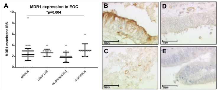
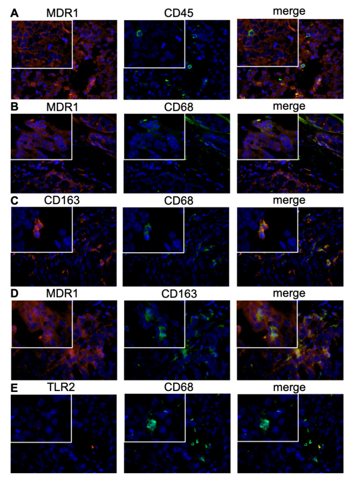
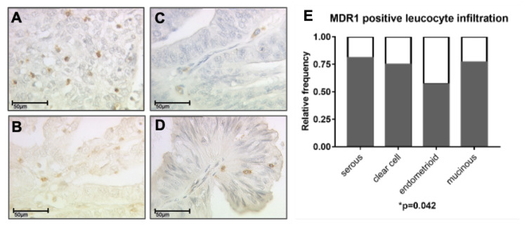
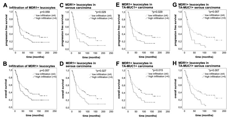
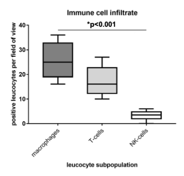
Multi drug resistance protein 1 (MDR1) expression on tumor cells has been widely investigated in context of drug resistance. However, the role of MDR1 on the immune cell infiltrate of solid tumors remains unknown. The aim of this study was to analyze the prognostic significance of a MDR1+ immune cell infiltrate in epithelial ovarian cancer (EOC) and to identify the MDR1+ leucocyte subpopulation. MDR1 expression was analyzed by immunohistochemistry in 156 EOC samples. In addition to MDR1+ cancer cells, we detected a MDR1+ leucocyte infiltrate (high infiltrate >4 leucocytes per field of view). Correlations and survival analyses were calculated. To identify immune cell subpopulations immunofluorescence double staining was performed. The MDR1+ leucocyte infiltrate was associated with human epidermal growth factor receptor 2 (HER2) (cc = 0.258, = 0.005) and tumor-associated mucin 1 (TA-MUC1) (cc = 0.202, = 0.022) expression on cancer cells. A high MDR1+ leucocyte infiltrate was associated with impaired survival, especially in patients whose carcinoma showed either serous histology (median OS 28.80 vs. 50.64 months, = 0.027, n = 91) or TA-MUC1 expression (median OS 30.60 vs. 63.36 months, = 0.015, n = 110). Similar findings for PFS suggest an influence of MDR1+ immune cells on the development of chemoresistance. A Cox regression analysis confirmed the independency of a high MDR1+ leucocyte infiltrate as prognostic factor. M2 macrophages were identified as main part of the MDR1+ leucocyte infiltrate expressing MDR1 as well as the M2 marker CD163 and the pan-macrophage marker CD68. Infiltration of MDR1+ leucocytes, mostly M2 macrophages, is associated with poor prognosis of EOC patients. Further understanding of the interaction of M2 macrophages, MDR1 and TA-MUC1 appears to be a key aspect to overcome chemoresistance in ovarian cancer.
多药耐药蛋白 1(MDR1)在肿瘤细胞中的表达已在药物耐药性方面得到广泛研究。然而,MDR1 对实体瘤中免疫细胞浸润的作用尚不清楚。本研究旨在分析上皮性卵巢癌(EOC)中 MDR1+免疫细胞浸润的预后意义,并鉴定 MDR1+白细胞亚群。采用免疫组织化学法分析了 156 例 EOC 样本中的 MDR1 表达。除了 MDR1+癌细胞,我们还检测到 MDR1+白细胞浸润(高浸润>每视野 4 个白细胞)。计算了相关性和生存分析。为了鉴定免疫细胞亚群,进行了免疫荧光双重染色。MDR1+白细胞浸润与人类表皮生长因子受体 2(HER2)(cc=0.258, =0.005)和肿瘤相关粘蛋白 1(TA-MUC1)(cc=0.202, =0.022)在癌细胞上的表达相关。高 MDR1+白细胞浸润与生存受损相关,尤其是在腺癌具有浆液性组织学(中位 OS 28.80 与 50.64 个月, =0.027,n=91)或 TA-MUC1 表达(中位 OS 30.60 与 63.36 个月, =0.015,n=110)的患者中。PFS 的相似发现提示 MDR1+免疫细胞对化疗耐药性的发展有影响。Cox 回归分析证实了高 MDR1+白细胞浸润作为预后因素的独立性。鉴定出 M2 巨噬细胞是 MDR1+白细胞浸润的主要部分,M2 巨噬细胞表达 MDR1 以及 M2 标志物 CD163 和泛巨噬细胞标志物 CD68。MDR1+白细胞浸润,主要是 M2 巨噬细胞,与 EOC 患者的预后不良相关。进一步了解 M2 巨噬细胞、MDR1 和 TA-MUC1 的相互作用似乎是克服卵巢癌化疗耐药性的关键方面。