Translational Tissue Engineering Center, Johns Hopkins University School of Medicine, Baltimore, MD, USA.
Department of Biomedical Engineering, Johns Hopkins University School of Medicine, Baltimore, MD, USA.
Sci Rep. 2020 May 20;10(1):8387. doi: 10.1038/s41598-020-65064-3.
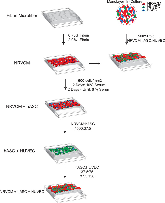
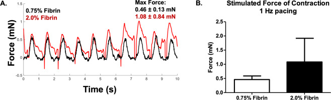
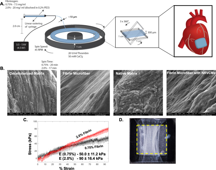
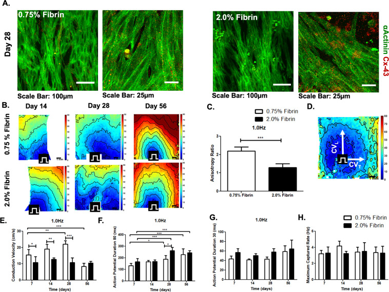
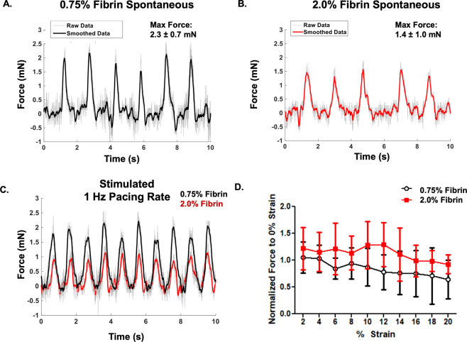
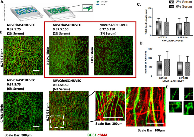
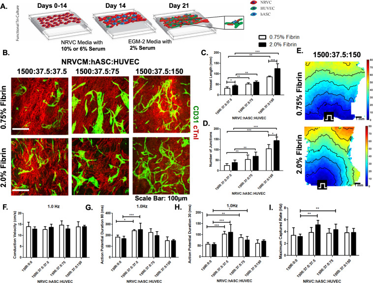
Cardiac tissue engineering strategies have the potential to regenerate functional myocardium following myocardial infarction. In this study, we utilized novel electrospun fibrin microfiber sheets of different stiffnesses (50.0 ± 11.2 kPa and 90.0 ± 16.4 kPa) to engineer biomimetic models of vascularized cardiac tissues. We characterized tissue assembly, electrophysiology, and contractility of neonatal rat ventricular cardiomyocytes (NRVCMs) cultured on these sheets. NRVCMs cultured on the softer substrates displayed higher conduction velocities (CVs) and improved electrophysiological properties. Human umbilical vein endothelial cells (HUVECs) formed dense networks on the sheets when co-cultured with human adipose-derived stem/stromal cells (hASCs). To achieve vascularized cardiac tissues, we tested various tri-culture protocols of NRVCM:hASC:HUVEC and found that a ratio of 1,500,000:37,500:150,000 cells/cm enabled the formation of robust endothelial networks while retaining statistically identical electrophysiological characteristics to NRVCM-only cultures. Tri-cultures at this ratio on 90 kPa substrates exhibited average CVs of 14 ± 0.6 cm/s, Action Potential Duration (APD)80 and APD30 of 152 ± 11 ms and 71 ± 6 ms, respectively, and maximum capture rate (MCR) of 3.9 ± 0.7 Hz. These data indicate the significant potential of generating densely packed endothelial networks together with electrically integrated cardiac cells in vitro as a physiologic 3D cardiac model.
心脏组织工程策略有可能在心肌梗死后再生功能性心肌。在这项研究中,我们利用不同硬度的新型静电纺丝纤维微片(50.0±11.2 kPa 和 90.0±16.4 kPa)来构建血管化心脏组织的仿生模型。我们对在这些薄片上培养的新生大鼠心室心肌细胞(NRVCM)的组织组装、电生理和收缩性进行了表征。在较软基底上培养的 NRVCM 表现出更高的传导速度(CV)和改善的电生理特性。当与人脂肪间充质干细胞(hASC)共培养时,人脐静脉内皮细胞(HUVEC)在薄片上形成密集的网络。为了实现血管化的心脏组织,我们测试了 NRVCM:hASC:HUVEC 的各种三培养方案,并发现 NRVCM:hASC:HUVEC 的比例为 1,500,000:37,500:150,000 个细胞/cm2 可以形成强大的内皮网络,同时保持与仅 NRVCM 培养相同的电生理特征。在 90 kPa 基质上以该比例进行的三培养显示出平均 CV 为 14±0.6 cm/s,动作电位持续时间(APD)80 和 APD30 分别为 152±11 ms 和 71±6 ms,最大捕获率(MCR)为 3.9±0.7 Hz。这些数据表明,在体外生成与电整合的心脏细胞一起形成密集的内皮网络具有显著的潜力,可作为生理 3D 心脏模型。