• 文献检索
  • 文档翻译
  • 深度研究
  • 学术资讯
  • Suppr Zotero 插件Zotero 插件
  • 邀请有礼
  • 套餐&价格
  • 历史记录
应用&插件
Suppr Zotero 插件Zotero 插件浏览器插件Mac 客户端Windows 客户端微信小程序
定价
高级版会员购买积分包购买API积分包
服务
文献检索文档翻译深度研究API 文档MCP 服务
关于我们
关于 Suppr公司介绍联系我们用户协议隐私条款
关注我们

Suppr 超能文献

核心技术专利:CN118964589B侵权必究
粤ICP备2023148730 号-1Suppr @ 2026

文献检索

告别复杂PubMed语法,用中文像聊天一样搜索,搜遍4000万医学文献。AI智能推荐,让科研检索更轻松。

立即免费搜索

文件翻译

保留排版,准确专业,支持PDF/Word/PPT等文件格式,支持 12+语言互译。

免费翻译文档

深度研究

AI帮你快速写综述,25分钟生成高质量综述,智能提取关键信息,辅助科研写作。

立即免费体验

在人工血液循环条件下对猪心进行的光学标测。

Optical mapping of the pig heart in situ under artificial blood circulation.

机构信息

Institute of Cardiology, Lithuanian University of Health Sciences, Kaunas, Lithuania.

Department of Cardiac, Thoracic and Vascular Surgery, Hospital of Lithuanian University of Health Sciences Kauno Klinikos, Lithuanian University of Health Sciences, Kaunas, Lithuania.

出版信息

Sci Rep. 2020 May 22;10(1):8548. doi: 10.1038/s41598-020-65464-5.

DOI:10.1038/s41598-020-65464-5
PMID:32444634
原文链接:https://pmc.ncbi.nlm.nih.gov/articles/PMC7244500/
Abstract

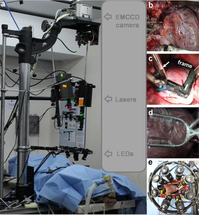
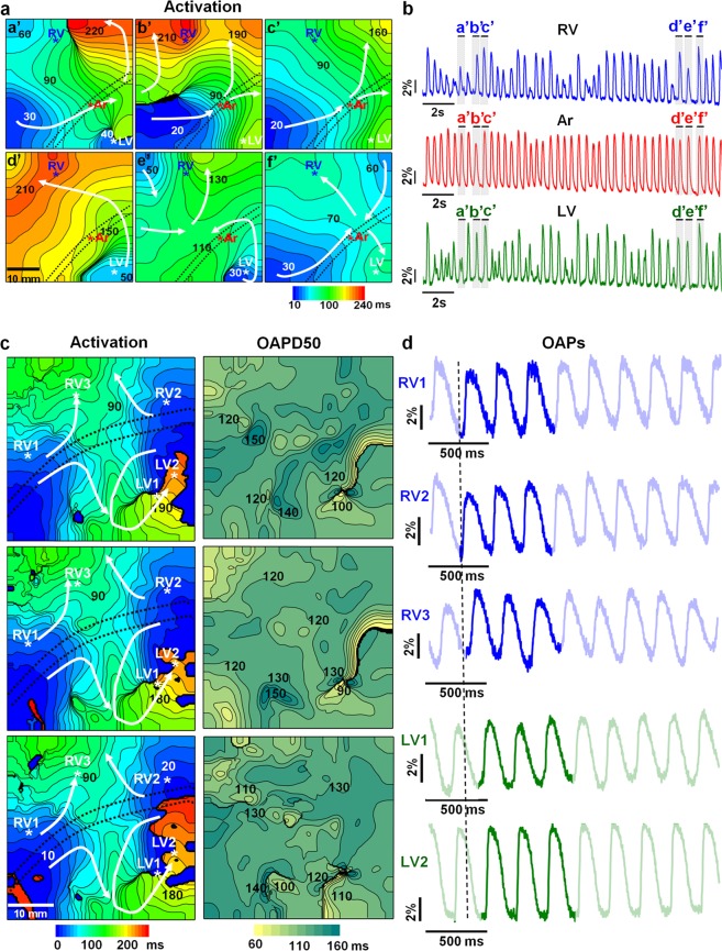
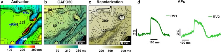
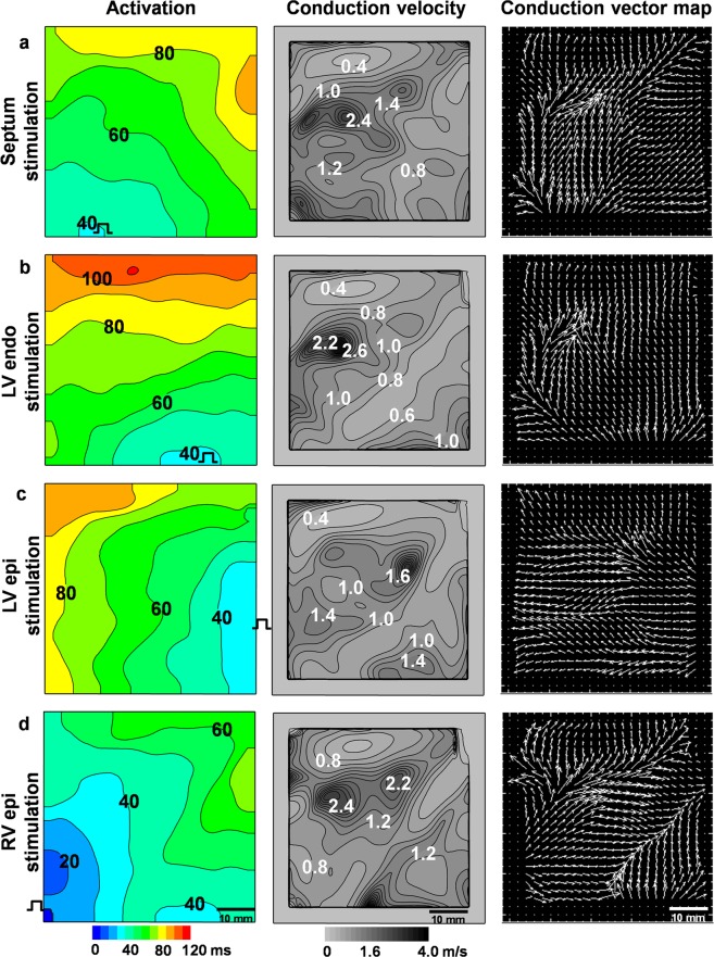
The emergence of optical imaging has revolutionized the investigation of cardiac electrical activity and associated disorders in various cardiac pathologies. The electrical signals of the heart and the propagation pathways are crucial for elucidating the mechanisms of various cardiac pathological conditions, including arrhythmia. The synthesis of near-infrared voltage-sensitive dyes and the voltage sensitivity of the FDA-approved dye Cardiogreen have increased the importance of optical mapping (OM) as a prospective tool in clinical practice. We aimed to develop a method for the high-spatiotemporal-resolution OM of the large animal hearts in situ using di-4-ANBDQBS and Cardiogreen under patho/physiological conditions. OM was adapted to monitor cardiac electrical behaviour in an open-chest pig heart model with physiological or artificial blood circulation. We detail the methods and display the OM data obtained using di-4-ANBDQBS and Cardiogreen. Activation time, action potential duration, repolarization time and conduction velocity maps were constructed. The technique was applied to track cardiac electrical activity during regional ischaemia and arrhythmia. Our study is the first to apply high-spatiotemporal-resolution OM in the pig heart in situ to record cardiac electrical activity qualitatively under artificial blood perfusion. The use of an FDA-approved voltage-sensitive dye and artificial blood perfusion in a swine model, which is generally accepted as a valuable pre-clinical model, demonstrates the promise of OM for clinical application.

摘要

光学成象的出现彻底改变了对各种心脏病理学中心脏电活动和相关疾病的研究。心脏的电信号和传播途径对于阐明各种心脏病理条件的机制至关重要,包括心律失常。近红外电压敏感染料的合成和 FDA 批准的染料 Cardiogreen 的电压敏感性增加了光学标测 (OM) 在临床实践中作为一种有前途的工具的重要性。我们旨在开发一种方法,用于在病理/生理条件下使用 di-4-ANBDQBS 和 Cardiogreen 对大动物心脏进行高时空分辨率的 OM。OM 被适当地用于监测具有生理或人工血液循环的开胸猪心模型中的心脏电行为。我们详细描述了方法,并显示了使用 di-4-ANBDQBS 和 Cardiogreen 获得的 OM 数据。构建了激活时间、动作电位持续时间、复极时间和传导速度图。该技术被应用于跟踪局部缺血和心律失常期间的心脏电活动。我们的研究首次应用高时空分辨率 OM 在猪心原位记录在人工血液灌注下的定性心脏电活动。在通常被认为是有价值的临床前模型的猪模型中使用 FDA 批准的电压敏感染料和人工血液灌注,表明 OM 在临床应用中的前景。

https://cdn.ncbi.nlm.nih.gov/pmc/blobs/26f4/7244500/a616e1bef67c/41598_2020_65464_Fig6_HTML.jpg
https://cdn.ncbi.nlm.nih.gov/pmc/blobs/26f4/7244500/972c212624e9/41598_2020_65464_Fig1_HTML.jpg
https://cdn.ncbi.nlm.nih.gov/pmc/blobs/26f4/7244500/71456a38046b/41598_2020_65464_Fig2_HTML.jpg
https://cdn.ncbi.nlm.nih.gov/pmc/blobs/26f4/7244500/18abb13b63bc/41598_2020_65464_Fig3_HTML.jpg
https://cdn.ncbi.nlm.nih.gov/pmc/blobs/26f4/7244500/ea82eff30796/41598_2020_65464_Fig4_HTML.jpg
https://cdn.ncbi.nlm.nih.gov/pmc/blobs/26f4/7244500/13e4c209275a/41598_2020_65464_Fig5_HTML.jpg
https://cdn.ncbi.nlm.nih.gov/pmc/blobs/26f4/7244500/a616e1bef67c/41598_2020_65464_Fig6_HTML.jpg

相似文献

1
Optical mapping of the pig heart in situ under artificial blood circulation.在人工血液循环条件下对猪心进行的光学标测。
Sci Rep. 2020 May 22;10(1):8548. doi: 10.1038/s41598-020-65464-5.
2
In vivo ratiometric optical mapping enables high-resolution cardiac electrophysiology in pig models.在体比率光学标测可实现猪模型的高分辨率心脏电生理学研究。
Cardiovasc Res. 2019 Sep 1;115(11):1659-1671. doi: 10.1093/cvr/cvz039.
3
Near-infrared voltage-sensitive fluorescent dyes optimized for optical mapping in blood-perfused myocardium.针对血液灌注心肌的光学映射优化的近红外电压敏感染料。
Heart Rhythm. 2007 Nov;4(11):1441-51. doi: 10.1016/j.hrthm.2007.07.012. Epub 2007 Jul 17.
4
Extracting surface activation time from the optically recorded action potential in three-dimensional myocardium.从三维心肌中的光记录动作电位中提取表面激活时间。
Biophys J. 2012 Jan 4;102(1):30-8. doi: 10.1016/j.bpj.2011.10.036. Epub 2012 Jan 3.
5
Evaluation of excitation propagation in the rabbit heart: optical mapping and transmural microelectrode recordings.兔心脏中兴奋传播的评估:光学标测与透壁微电极记录
PLoS One. 2015 Apr 16;10(4):e0123050. doi: 10.1371/journal.pone.0123050. eCollection 2015.
6
Voltage-Sensitive Fluorescence of Indocyanine Green in the Heart.心脏中吲哚菁绿的电压敏感性荧光
Biophys J. 2016 Feb 2;110(3):723-732. doi: 10.1016/j.bpj.2015.12.021.
7
Optical mapping at increased illumination intensities.在增加光照强度下的光学作图。
J Biomed Opt. 2012 Sep;17(9):96007-1. doi: 10.1117/1.JBO.17.9.096007.
8
Optical Mapping Approaches on Muscleblind-Like Compound Knockout Mice for Understanding Mechanistic Insights Into Ventricular Arrhythmias in Myotonic Dystrophy.利用类肌肉盲复合基因敲除小鼠的光学映射方法来深入了解强直性肌营养不良中心室心律失常的机制。
J Am Heart Assoc. 2017 Apr 17;6(4):e005191. doi: 10.1161/JAHA.116.005191.
9
Construction of 3D MR image-based computer models of pathologic hearts, augmented with histology and optical fluorescence imaging to characterize action potential propagation.构建基于三维磁共振成像的病理性心脏计算机模型,并结合组织学和光学荧光成像来描述动作电位的传播。
Med Image Anal. 2012 Feb;16(2):505-23. doi: 10.1016/j.media.2011.11.007. Epub 2011 Dec 6.
10
Cardiac Optical Mapping in Situ in Swine Models: A View of the Current Situation.猪模型原位心脏光学映射:现状观察
Medicina (Kaunas). 2020 Nov 18;56(11):620. doi: 10.3390/medicina56110620.

引用本文的文献

1
Optical mapping and optogenetics in cardiac electrophysiology research and therapy: a state-of-the-art review.光学绘图与光遗传学在心脏电生理学研究与治疗中的应用:最新综述。
Europace. 2024 Feb 1;26(2). doi: 10.1093/europace/euae017.
2
KairoSight: Open-Source Software for the Analysis of Cardiac Optical Data Collected From Multiple Species.KairoSight:用于分析从多个物种收集的心脏光学数据的开源软件。
Front Physiol. 2021 Oct 29;12:752940. doi: 10.3389/fphys.2021.752940. eCollection 2021.
3
Stop the beat to see the rhythm: excitation-contraction uncoupling in cardiac research.

本文引用的文献

1
Sympathetic Nervous Regulation of Calcium and Action Potential Alternans in the Intact Heart.完整心脏中交感神经对钙及动作电位交替的调节
Front Physiol. 2018 Jan 23;9:16. doi: 10.3389/fphys.2018.00016. eCollection 2018.
2
Spectral characteristics of voltage-sensitive indocyanine green fluorescence in the heart.心脏中电压敏感性吲哚菁绿荧光的光谱特征。
Sci Rep. 2017 Aug 11;7(1):7983. doi: 10.1038/s41598-017-08168-7.
3
Cardiac sympathetic innervation via middle cervical and stellate ganglia and antiarrhythmic mechanism of bilateral stellectomy.
停止跳动以观察节奏:心脏研究中的兴奋-收缩脱偶。
Am J Physiol Heart Circ Physiol. 2021 Dec 1;321(6):H1005-H1013. doi: 10.1152/ajpheart.00477.2021. Epub 2021 Oct 8.
4
Cardiac Optical Mapping in Situ in Swine Models: A View of the Current Situation.猪模型原位心脏光学映射:现状观察
Medicina (Kaunas). 2020 Nov 18;56(11):620. doi: 10.3390/medicina56110620.
通过颈中神经节和星状神经节的心脏交感神经支配以及双侧星状神经节切除术的抗心律失常机制。
Am J Physiol Heart Circ Physiol. 2017 Mar 1;312(3):H392-H405. doi: 10.1152/ajpheart.00644.2016. Epub 2016 Dec 23.
4
Optical Mapping of Membrane Potential and Epicardial Deformation in Beating Hearts.跳动心脏中膜电位和心外膜变形的光学映射
Biophys J. 2016 Jul 26;111(2):438-451. doi: 10.1016/j.bpj.2016.03.043.
5
Voltage-Sensitive Fluorescence of Indocyanine Green in the Heart.心脏中吲哚菁绿的电压敏感性荧光
Biophys J. 2016 Feb 2;110(3):723-732. doi: 10.1016/j.bpj.2015.12.021.
6
Optical Imaging of Cardiac Action Potential.心脏动作电位的光学成像
Adv Exp Med Biol. 2015;859:299-311. doi: 10.1007/978-3-319-17641-3_12.
7
Evaluation of excitation propagation in the rabbit heart: optical mapping and transmural microelectrode recordings.兔心脏中兴奋传播的评估:光学标测与透壁微电极记录
PLoS One. 2015 Apr 16;10(4):e0123050. doi: 10.1371/journal.pone.0123050. eCollection 2015.
8
Simulating photon scattering effects in structurally detailed ventricular models using a Monte Carlo approach.使用蒙特卡罗方法在结构详细的心室模型中模拟光子散射效应。
Front Physiol. 2014 Sep 9;5:338. doi: 10.3389/fphys.2014.00338. eCollection 2014.
9
Real-time imaging of electrical signals with an infrared FDA-approved dye.使用经美国食品药品监督管理局批准的红外染料对电信号进行实时成像。
Biophys J. 2014 Sep 16;107(6):L09-12. doi: 10.1016/j.bpj.2014.07.054.
10
Small and large animal models in cardiac contraction research: advantages and disadvantages.心脏收缩研究中小动物模型的优缺点。
Pharmacol Ther. 2014 Mar;141(3):235-49. doi: 10.1016/j.pharmthera.2013.10.007. Epub 2013 Oct 15.