Biomedical Research Imaging Center, Chapel Hill, NC, USA; Department of Radiology, University of North Carolina at Chapel Hill, Chapel Hill, NC, USA.
Biomedical Research Imaging Center, Chapel Hill, NC, USA; Department of Radiology, University of North Carolina at Chapel Hill, Chapel Hill, NC, USA.
Neuroimage. 2020 Sep;218:116978. doi: 10.1016/j.neuroimage.2020.116978. Epub 2020 May 21.
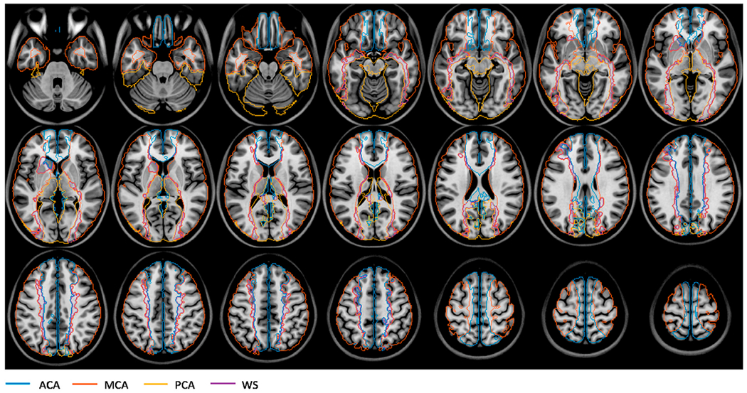
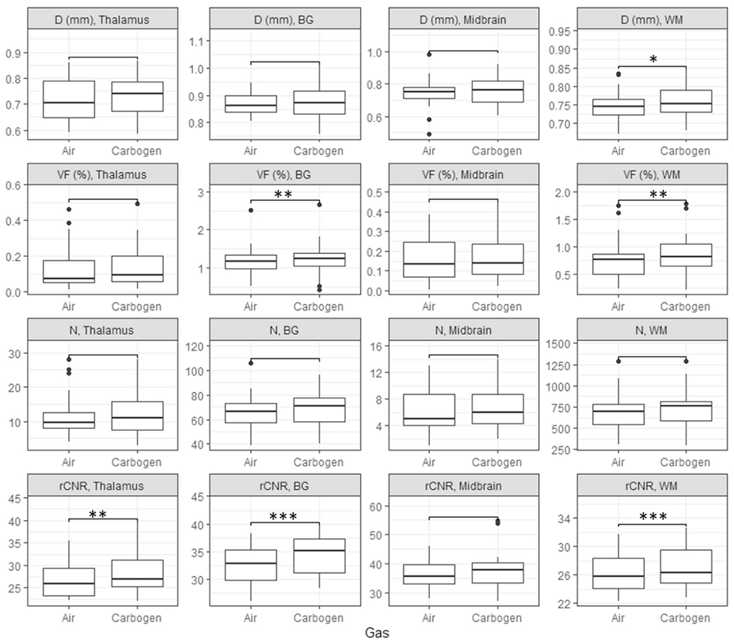
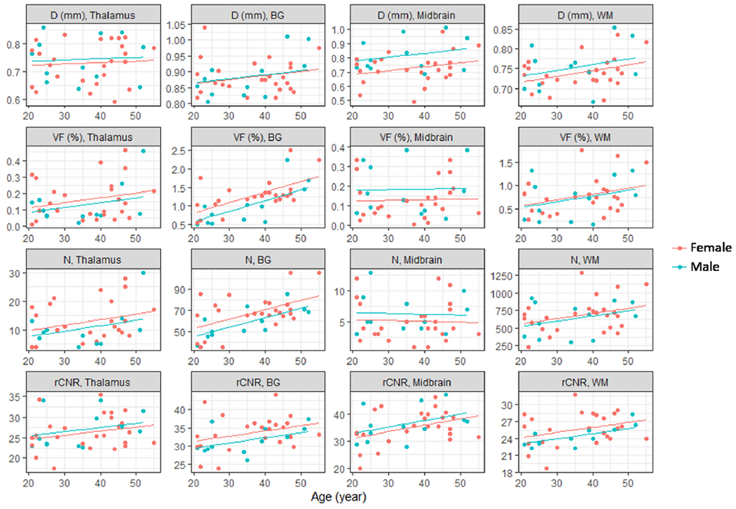
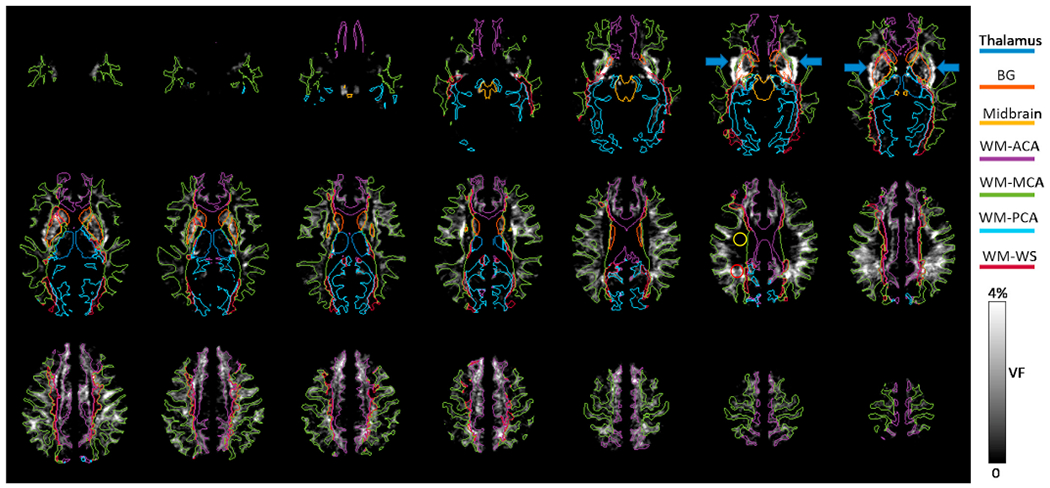
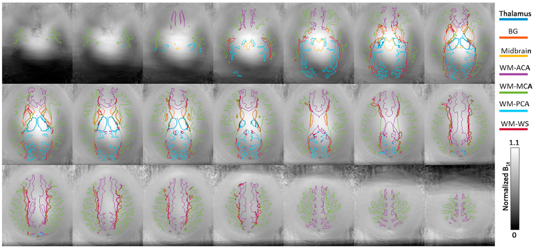
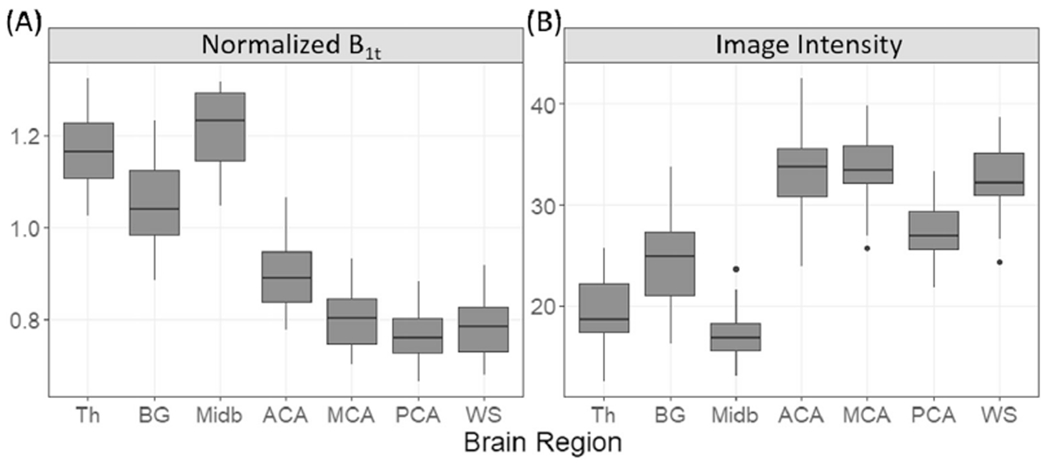
Perivascular spaces (PVSs) are fluid-filled spaces surrounding penetrating blood vessels in the brain and are an integral pathway of the glymphatic system. A PVS and the enclosed blood vessel are commonly visualized as a single vessel-like complex (denoted as PVSV) in high-resolution MRI images. Quantitative characterization of the PVSV morphology in MRI images in healthy subjects may serve as a reference for detecting disease related PVS and/or blood vessel alterations in patients with brain diseases. To this end, we evaluated the age dependences, spatial heterogeneities, and dynamic properties of PVSV morphological features in 45 healthy subjects (21-55 years old), using an ultra-high-resolution three-dimensional transverse relaxation time weighted MRI sequence (0.41 × 0.41 × 0.4 mm) at 7T. Quantitative PVSV parameters, including apparent diameter, count, volume fraction (VF), and relative contrast to noise ratio (rCNR) were calculated in the white matter and subcortical structures. Dynamic changes were induced by carbogen breathing which are known to induce vasodilation and increase the blood oxygenation level in the brain. PVSV count and VF significantly increased with age in basal ganglia (BG), so did rCNR in BG, midbrain, and white matter (WM). Apparent PVSV diameter also showed a positive association with age in the three brain regions, although it did not reach statistical significance. The PVSV VF and count showed large inter-subject variations, with coefficients of variation ranging from 0.17 to 0.74 after regressing out age and gender effects. Both apparent diameter and VF exhibited significant spatial heterogeneity, which cannot be explained solely by radio-frequency field inhomogeneities. Carbogen breathing significantly increased VF in BG and WM, and rCNR in thalamus, BG, and WM compared to air breathing. Our results are consistent with gradual dilation of PVSs with age in healthy adults. The PVSV morphology exhibited spatial heterogeneity and large inter-subject variations and changed during carbogen breathing compared to air breathing.
血管周围间隙(PVS)是围绕脑内穿透性血管的充满液体的空间,是脑内淋巴系统的重要途径。在高分辨率 MRI 图像中,PVS 和包含的血管通常被视为单一的血管样复合体(表示为 PVSV)。在健康受试者的 MRI 图像中对 PVSV 形态的定量特征进行描述,可作为检测脑疾病患者与疾病相关的 PVS 和/或血管改变的参考。为此,我们使用 7T 超高分辨率三维横向弛豫时间加权 MRI 序列(0.41×0.41×0.4mm)对 45 名健康受试者(21-55 岁)进行了研究,评估了 PVSV 形态特征的年龄依赖性、空间异质性和动态特性。在白质和皮质下结构中计算了表观直径、计数、体积分数(VF)和相对对比噪声比(rCNR)等定量 PVSV 参数。已知碳酸氧合呼吸会引起血管扩张并增加大脑中的血氧水平,从而引起动态变化。在基底节(BG)、中脑和白质(WM)中,PVSV 计数和 VF 随年龄显著增加,BG、中脑和 WM 的 rCNR 也随之增加。尽管在三个脑区中,表观 PVSV 直径与年龄之间也存在正相关关系,但并未达到统计学意义。PVSV VF 和计数表现出较大的个体间变异性,在回归年龄和性别效应后,变异系数范围为 0.17 至 0.74。表观直径和 VF 均表现出明显的空间异质性,这不能仅用射频场不均匀性来解释。与空气呼吸相比,碳酸氧合呼吸显著增加了 BG 和 WM 中的 VF,以及丘脑、BG 和 WM 中的 rCNR。我们的结果与健康成年人的 PVS 随年龄逐渐扩张一致。与空气呼吸相比,PVSV 形态在碳酸氧合呼吸期间表现出空间异质性和较大的个体间变异性,并发生变化。