• 文献检索
  • 文档翻译
  • 深度研究
  • 学术资讯
  • Suppr Zotero 插件Zotero 插件
  • 邀请有礼
  • 套餐&价格
  • 历史记录
应用&插件
Suppr Zotero 插件Zotero 插件浏览器插件Mac 客户端Windows 客户端微信小程序
定价
高级版会员购买积分包购买API积分包
服务
文献检索文档翻译深度研究API 文档MCP 服务
关于我们
关于 Suppr公司介绍联系我们用户协议隐私条款
关注我们

Suppr 超能文献

核心技术专利:CN118964589B侵权必究
粤ICP备2023148730 号-1Suppr @ 2026

文献检索

告别复杂PubMed语法,用中文像聊天一样搜索,搜遍4000万医学文献。AI智能推荐,让科研检索更轻松。

立即免费搜索

文件翻译

保留排版,准确专业,支持PDF/Word/PPT等文件格式,支持 12+语言互译。

免费翻译文档

深度研究

AI帮你快速写综述,25分钟生成高质量综述,智能提取关键信息,辅助科研写作。

立即免费体验

微血管手术中电子内窥镜与手术显微镜的比较:一项模拟非劣效性试验。

The Exoscope versus operating microscope in microvascular surgery: A simulation non-inferiority trial.

作者信息

Pafitanis Georgios, Hadjiandreou Michalis, Alamri Alexander, Uff Christopher, Walsh Daniel, Myers Simon

机构信息

Group for Academic Plastic Surgery, Microvascular Anastomosis Simulation Hub, The Blizard Institute, Queen Mary University of London, London, UK.

Department of Plastic Surgery, Great Ormond Street Hospital for Children Foundation Trust, London, UK.

出版信息

Arch Plast Surg. 2020 May;47(3):242-249. doi: 10.5999/aps.2019.01473. Epub 2020 May 15.

DOI:10.5999/aps.2019.01473
PMID:32453933
原文链接:https://pmc.ncbi.nlm.nih.gov/articles/PMC7264907/
Abstract

BACKGROUND

The Exoscope is a novel high-definition digital camera system. There is limited evidence signifying the use of exoscopic devices in microsurgery. This trial objectively assesses the effects of the use of the Exoscope as an alternative to the standard operating microscope (OM) on the performance of experts in a simulated microvascular anastomosis.

METHODS

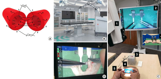
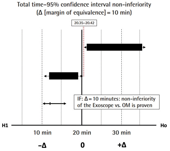
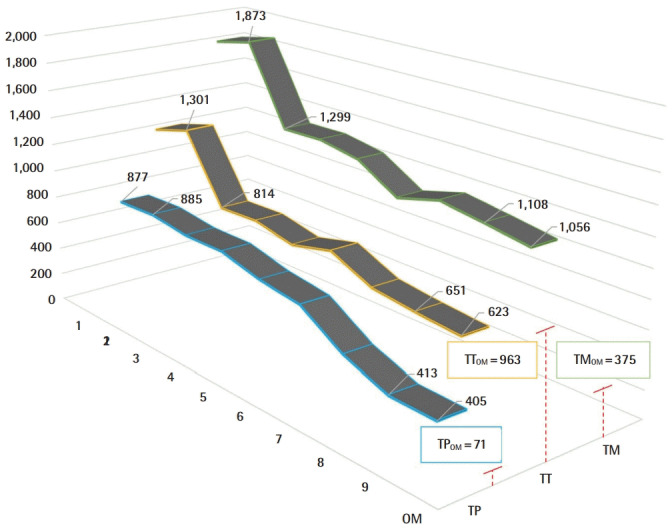
Modus V Exoscope and OM were used by expert microsurgeons to perform standardized tasks. Hand-motion analyzer measured the total pathlength (TP), total movements (TM), total time (TT), and quality of end-product anastomosis. A clinical margin of TT was performed to prove non-inferiority. An expert performed consecutive microvascular anastomoses to provide the exoscopic learning curve until reached plateau in TT.

RESULTS

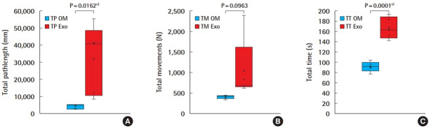
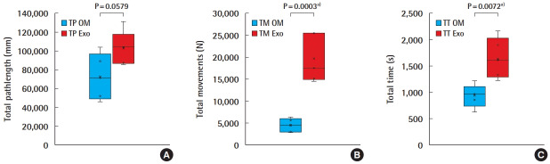
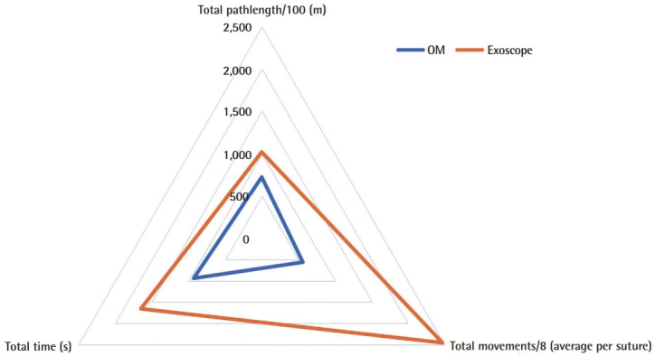
Ten micro sutures and 10 anastomoses were performed. Analysis demonstrated statistically significant differences in performing micro sutures for TP, TM, and TT. There was statistical significance in TM and TT, however, marginal non-significant difference in TP regarding microvascular anastomoses performance. The intimal suture line analysis demonstrated no statistically significant differences. Non-inferiority results based on clinical inferiority margin (Δ) of TT=10 minutes demonstrated an absolute difference of 0.07 minutes between OM and Exoscope cohorts. A 51%, 58%, and 46% improvement or reduction was achieved in TT, TM, TP, respectively, during the exoscopic microvascular anastomosis learning curve.

CONCLUSIONS

This study demonstrated that experts' Exoscope anastomoses appear non-inferior to the OM anastomoses. Exoscopic microvascular anastomosis was more time consuming but end-product (patency) in not clinically inferior. Experts' "warm-up" learning curve is steep but swift and may prove to reach clinical equality.

摘要

背景

外视镜是一种新型高清数码相机系统。关于在显微手术中使用外视镜设备的证据有限。本试验客观评估了使用外视镜替代标准手术显微镜(OM)对模拟微血管吻合术中专家操作的影响。

方法

显微外科专家使用Modus V外视镜和OM执行标准化任务。手动运动分析仪测量总路径长度(TP)、总运动次数(TM)、总时间(TT)以及最终产品吻合的质量。进行TT的临床差值分析以证明非劣效性。一名专家连续进行微血管吻合以提供外视镜学习曲线,直至TT达到平稳状态。

结果

进行了10次显微缝合和10次吻合。分析表明,在进行显微缝合时,TP、TM和TT存在统计学显著差异。在TM和TT方面存在统计学意义,但在微血管吻合操作的TP方面存在边缘性非显著差异。内膜缝合线分析显示无统计学显著差异。基于TT = 10分钟的临床劣效性差值(Δ)的非劣效性结果表明,OM组和外视镜组之间的绝对差值为0.07分钟。在外视镜微血管吻合学习曲线期间,TT、TM、TP分别提高或降低了51%、58%和46%。

结论

本研究表明,专家使用外视镜进行的吻合似乎不劣于使用OM进行的吻合。外视镜微血管吻合耗时更长,但最终产品(通畅率)在临床上并不逊色。专家的“热身”学习曲线陡峭但迅速,可能会达到临床等效性。

https://cdn.ncbi.nlm.nih.gov/pmc/blobs/d0c9/7264907/07d2b559e6a2/aps-2019-01473f6.jpg
https://cdn.ncbi.nlm.nih.gov/pmc/blobs/d0c9/7264907/40da4b1fa2d5/aps-2019-01473f1.jpg
https://cdn.ncbi.nlm.nih.gov/pmc/blobs/d0c9/7264907/383ac3e9d246/aps-2019-01473f2.jpg
https://cdn.ncbi.nlm.nih.gov/pmc/blobs/d0c9/7264907/060c69a1eb74/aps-2019-01473f3.jpg
https://cdn.ncbi.nlm.nih.gov/pmc/blobs/d0c9/7264907/4549136b5498/aps-2019-01473f4.jpg
https://cdn.ncbi.nlm.nih.gov/pmc/blobs/d0c9/7264907/7ad35a167dc9/aps-2019-01473f5.jpg
https://cdn.ncbi.nlm.nih.gov/pmc/blobs/d0c9/7264907/07d2b559e6a2/aps-2019-01473f6.jpg

相似文献

1
The Exoscope versus operating microscope in microvascular surgery: A simulation non-inferiority trial.微血管手术中电子内窥镜与手术显微镜的比较:一项模拟非劣效性试验。
Arch Plast Surg. 2020 May;47(3):242-249. doi: 10.5999/aps.2019.01473. Epub 2020 May 15.
2
Microsurgical training using an model: microscope 3D exoscope.使用 模型进行显微外科训练:显微镜 3D 外窥镜。
Acta Otorhinolaryngol Ital. 2022 Jun;42(3):223-229. doi: 10.14639/0392-100X-N1946.
3
A high-definition 3D exoscope as an alternative to the operating microscope in spinal microsurgery.高清3D外视镜在脊柱显微手术中可替代手术显微镜。
J Neurosurg Spine. 2020 Jul 10;33(5):705-714. doi: 10.3171/2020.4.SPINE20374. Print 2020 Nov 1.
4
3D Exoscope-Assisted Microvascular Anastomosis: An Evaluation on Latex Vessel Models.3D 电子内窥镜辅助微血管吻合术:对乳胶血管模型的评估
J Clin Med. 2020 Oct 21;9(10):3373. doi: 10.3390/jcm9103373.
5
Microvascular anastomosis under 3D exoscope or endoscope magnification: A proof-of-concept study.3D 外视镜或内镜放大下的微血管吻合术:一项概念验证研究。
Surg Neurol Int. 2018 Jun 4;9:115. doi: 10.4103/sni.sni_36_18. eCollection 2018.
6
Advancement of Surgical Visualization Methods: Comparison Study Between Traditional Microscopic Surgery and a Novel Robotic Optoelectronic Visualization Tool for Spinal Surgery.手术可视化方法的进展:传统显微手术与新型脊柱手术机器人光电可视化工具的比较研究
World Neurosurg. 2017 Feb;98:273-277. doi: 10.1016/j.wneu.2016.11.003. Epub 2016 Nov 9.
7
Even short-term training improves the skills of novice exoscope users: a prospective laboratory experiment.即使是短期培训也能提高新手外骨骼使用者的技能:一项前瞻性实验室实验。
Acta Neurochir (Wien). 2024 Mar 1;166(1):118. doi: 10.1007/s00701-024-05975-6.
8
Use of the 4K-3D ORBEYE Exoscope for Supermicrosurgical Lymphaticovenular Anastomosis: A Case Report.使用4K-3D ORBEYE电子外视镜进行超显微淋巴管静脉吻合术:一例报告
Cureus. 2024 Oct 10;16(10):e71198. doi: 10.7759/cureus.71198. eCollection 2024 Oct.
9
Microvascular anastomotic coupler application learning curve: A curriculum supporting further deliberate practice in ex-vivo simulation models.微血管吻合器应用学习曲线:支持进一步在体外模拟模型中进行刻意练习的课程。
J Plast Reconstr Aesthet Surg. 2019 Feb;72(2):203-210. doi: 10.1016/j.bjps.2018.10.021. Epub 2018 Nov 2.
10
A Novel Use of the Exoscope for In-Vivo Microvascular Free Flaps.外视镜在体内微血管游离皮瓣中的新应用。
J Oral Maxillofac Surg. 2023 Apr;81(4):511-514. doi: 10.1016/j.joms.2022.12.013. Epub 2022 Dec 29.

引用本文的文献

1
Enhancing submandibular gland resection: A retrospective study on the efficacy of the ORBEYE 3D exoscope.提高下颌下腺切除术效果:一项关于ORBEYE 3D电子内窥镜疗效的回顾性研究
Oral Maxillofac Surg. 2025 Sep 10;29(1):149. doi: 10.1007/s10006-025-01446-z.
2
Comparison of operative microscope and exoscope for execution of microanastomoses on an artificial model.在人工模型上使用手术显微镜和外视镜进行显微吻合术的比较。
Front Surg. 2025 Aug 8;12:1573333. doi: 10.3389/fsurg.2025.1573333. eCollection 2025.
3
Exoscopes in Neurosurgery: A Bibliometric and Visualization Analysis.

本文引用的文献

1
The Exoscope in Neurosurgery: An Innovative "Point of View". A Systematic Review of the Technical, Surgical, and Educational Aspects.神经外科手术中的外视镜:一种创新的“视角”。对技术、手术及教育方面的系统评价
World Neurosurg. 2019 Apr;124:136-144. doi: 10.1016/j.wneu.2018.12.202. Epub 2019 Jan 17.
2
Potential of performing a microvascular free flap reconstruction using solely a 3D exoscope instead of a conventional microscope.仅使用3D外视镜而非传统显微镜进行微血管游离皮瓣重建的可能性。
J Plast Reconstr Aesthet Surg. 2018 Nov;71(11):1664-1678. doi: 10.1016/j.bjps.2018.07.032. Epub 2018 Aug 8.
3
Preclinical Experience Using a New Robotic System Created for Microsurgery.
神经外科中的外视镜:文献计量学与可视化分析
Asian J Neurosurg. 2025 Mar 31;20(3):437-447. doi: 10.1055/s-0045-1806860. eCollection 2025 Sep.
4
Fluorescence-Guided Surgery for Gliomas: Past, Present, and Future.胶质瘤的荧光引导手术:过去、现在与未来
Cancers (Basel). 2025 May 30;17(11):1837. doi: 10.3390/cancers17111837.
5
Learning Curve and Ergonomics Associated with the 3D-monitor-assisted Microsurgery Using a Digital Microscope.使用数字显微镜的3D显示器辅助显微手术的学习曲线和人体工程学
J Plast Reconstr Surg. 2022 Jun 17;2(1):1-8. doi: 10.53045/jprs.2021-0026. eCollection 2023 Jan 27.
6
Post-operative monitoring of tissue perfusion in murine skin flaps using enhanced thermal imaging.使用增强型热成像技术对小鼠皮瓣组织灌注进行术后监测。
Biomed Opt Express. 2025 Mar 11;16(4):1406-1422. doi: 10.1364/BOE.551057. eCollection 2025 Apr 1.
7
Application of the ORBEYE Three-Dimensional Exoscope for Parotidectomies.ORBEYE三维外视镜在腮腺切除术中的应用。
J Clin Med. 2024 Dec 26;14(1):47. doi: 10.3390/jcm14010047.
8
From Microscopic to Exoscopic Microsurgery: Are We Facing a Change of Paradigm?从微观到宏观显微镜手术:我们是否面临范式转变?
Adv Tech Stand Neurosurg. 2024;53:27-49. doi: 10.1007/978-3-031-67077-0_3.
9
Microscopic subinguinal varicocelectomy with video telescopic operating microscope (VITOM) telescope: outcome analysis.使用视频伸缩式手术显微镜(VITOM)望远镜进行显微镜下腹股沟下精索静脉结扎术:结果分析
Transl Androl Urol. 2024 Apr 30;13(4):560-567. doi: 10.21037/tau-23-586. Epub 2024 Apr 17.
10
Even short-term training improves the skills of novice exoscope users: a prospective laboratory experiment.即使是短期培训也能提高新手外骨骼使用者的技能:一项前瞻性实验室实验。
Acta Neurochir (Wien). 2024 Mar 1;166(1):118. doi: 10.1007/s00701-024-05975-6.
用于微创手术的新型机器人系统的临床前经验。
Plast Reconstr Surg. 2018 Nov;142(5):1367-1376. doi: 10.1097/PRS.0000000000004939.
4
First Clinical Applications of a High-Definition Three-Dimensional Exoscope in Pediatric Neurosurgery.高清三维外视镜在小儿神经外科的首次临床应用
Cureus. 2018 Jan 24;10(1):e2108. doi: 10.7759/cureus.2108.
5
Initial Experience Using a High-Definition 3-Dimensional Exoscope System for Microneurosurgery.高清三维手术显微镜系统在微创神经外科中的初步应用经验
Oper Neurosurg (Hagerstown). 2018 Apr 1;14(4):395-401. doi: 10.1093/ons/opx145.
6
VITOM 3D: Preliminary Experience in Cranial Surgery.VITOM 3D:颅脑手术的初步经验
World Neurosurg. 2017 Nov;107:663-668. doi: 10.1016/j.wneu.2017.08.083. Epub 2017 Aug 24.
7
Vitom-3D for Exoscopic Neurosurgery: Initial Experience in Cranial and Spinal Procedures.用于内镜神经外科手术的Vitom-3D:颅脑和脊柱手术的初步经验。
World Neurosurg. 2017 Sep;105:153-162. doi: 10.1016/j.wneu.2017.05.109. Epub 2017 May 27.
8
From Exoscope into the Next Generation.从外视镜迈向新一代。
J Korean Neurosurg Soc. 2017 May;60(3):289-293. doi: 10.3340/jkns.2017.0202.003. Epub 2017 May 1.
9
Comprehensive Analysis of Chicken Vessels as Microvascular Anastomosis Training Model.鸡血管作为微血管吻合训练模型的综合分析
Arch Plast Surg. 2017 Jan;44(1):12-18. doi: 10.5999/aps.2017.44.1.12. Epub 2017 Jan 20.
10
Advancement of Surgical Visualization Methods: Comparison Study Between Traditional Microscopic Surgery and a Novel Robotic Optoelectronic Visualization Tool for Spinal Surgery.手术可视化方法的进展:传统显微手术与新型脊柱手术机器人光电可视化工具的比较研究
World Neurosurg. 2017 Feb;98:273-277. doi: 10.1016/j.wneu.2016.11.003. Epub 2016 Nov 9.