Kang Bo Young, Jeon Byung-Joon, Lee Kyeong-Tae, Mun Goo-Hyun
Department of Plastic Surgery, Samsung Medical Center, Sungkyunkwan University School of Medicine, Seoul, Korea.
Arch Plast Surg. 2017 Jan;44(1):12-18. doi: 10.5999/aps.2017.44.1.12. Epub 2017 Jan 20.
Nonliving chickens are commonly used as a microvascular anastomosis training model. However, previous studies have investigated only a few types of vessel, and no study has compared the characteristics of the various vessels. The present study evaluated the anatomic characteristics of various chicken vessels as a training model.
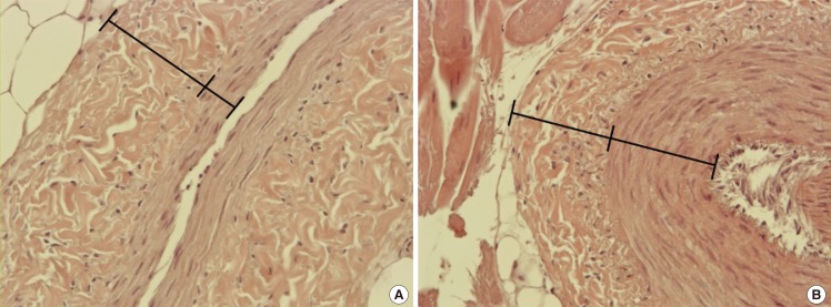
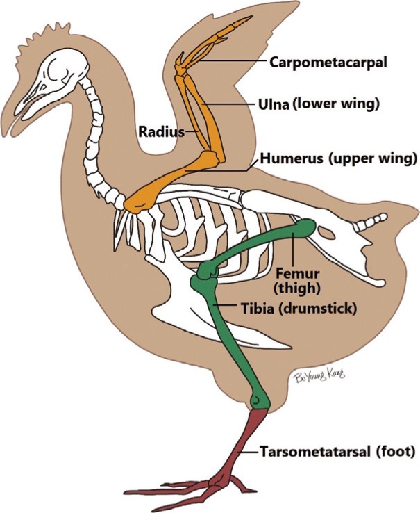
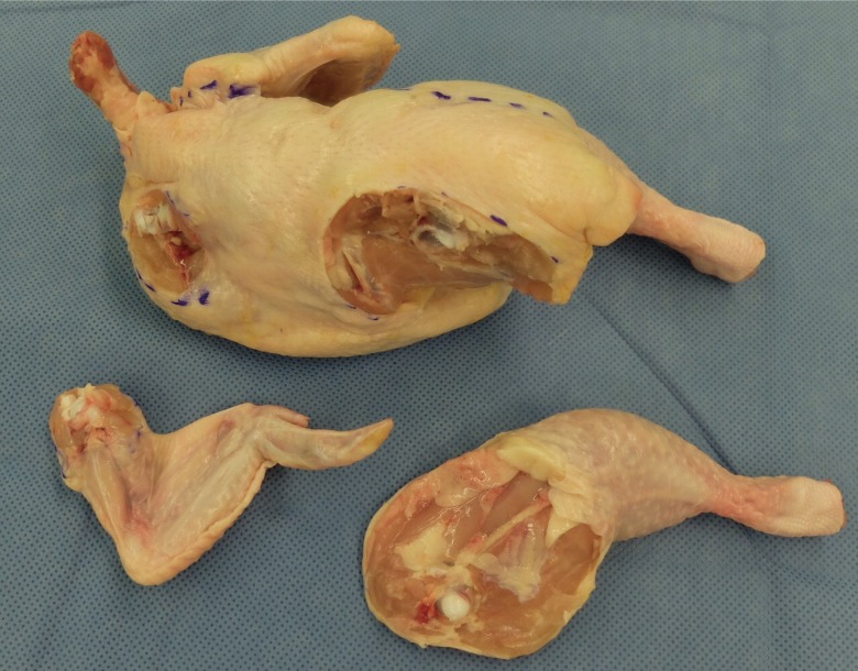
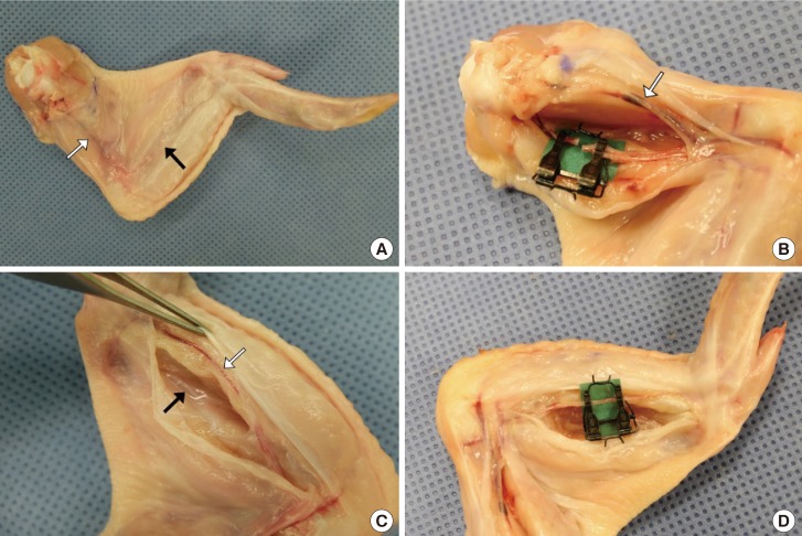
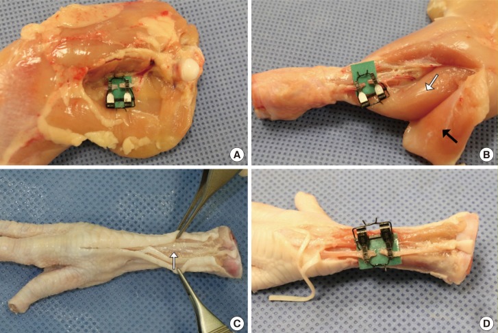
Eight vessels-the brachial artery, basilic vein, radial artery, ulnar artery, ischiatic artery and vein, cranial tibial artery, and common dorsal metatarsal artery-were evaluated in 26 fresh chickens and 30 chicken feet for external diameter (ED) and thicknesses of the tunica adventitia and media. The dissection time from skin incision to application of vessel clamps was also measured.
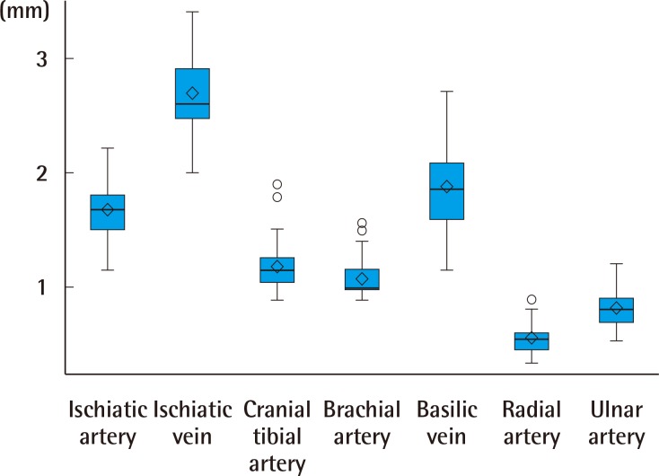
The EDs of the vessels varied. The ischiatic vein had the largest ED of 2.69±0.33 mm, followed by the basilic vein (1.88±0.36 mm), ischiatic artery (1.68±0.24 mm), common dorsal metatarsal artery (1.23±0.23 mm), cranial tibial artery (1.18±0.19 mm), brachial artery (1.08±0.15 mm), ulnar artery (0.82±0.13 mm), and radial artery (0.56±0.12 mm), and the order of size was consistent across all subjects. Thicknesses of the tunica adventitia and media were also diverse, ranging from 74.09±19.91 µm to 158.66±40.25 µm (adventitia) and from 31.2±7.13 µm to 154.15±46.48 µm (media), respectively. Mean dissection time was <3 minutes for all vessels.
Our results suggest that nonliving chickens can provide various vessels with different anatomic characteristics, which can allow trainees the choice of an appropriate microvascular anastomosis training model depending on their purpose and skillfulness.
死鸡常用于微血管吻合训练模型。然而,以往的研究仅调查了少数几种血管类型,尚无研究比较各种血管的特征。本研究评估了各种鸡血管作为训练模型的解剖学特征。
在26只新鲜鸡和30只鸡脚上评估了8种血管——肱动脉、贵要静脉、桡动脉、尺动脉、坐骨动脉和静脉、胫前动脉以及跖背总动脉——的外径(ED)以及外膜和中膜的厚度。还测量了从皮肤切开到应用血管夹的解剖时间。
各血管的外径各不相同。坐骨静脉的外径最大,为2.69±0.33毫米,其次是贵要静脉(1.88±0.36毫米)、坐骨动脉(1.68±0.24毫米)、跖背总动脉(1.23±0.23毫米)、胫前动脉(1.18±0.19毫米)、肱动脉(1.08±0.15毫米)、尺动脉(0.82±0.13毫米)和桡动脉(0.56±0.12毫米),且所有受试者的大小顺序一致。外膜和中膜的厚度也各不相同,分别为74.09±19.91微米至158.66±40.25微米(外膜)和31.2±7.13微米至154.15±46.48微米(中膜)。所有血管的平均解剖时间均小于3分钟。
我们的结果表明,死鸡可提供具有不同解剖学特征的各种血管,这能让受训者根据自身目的和熟练程度选择合适的微血管吻合训练模型。