Vasiliadis Konstantinos, Moschou Elena, Papaioannou Sofia, Tzitzis Panagiotis, Totsi Albion, Dimou Stamatia, Lazaridou Eleni, Kapetanos Dimitrios, Papavasiliou Christos
First Surgical Department, Thessaloniki, Greece.
Department of Radiology, General Hospital Papageorgiou, Thessaloniki, Greece.
Ann Hepatobiliary Pancreat Surg. 2020 May 31;24(2):221-227. doi: 10.14701/ahbps.2020.24.2.221.
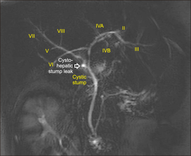
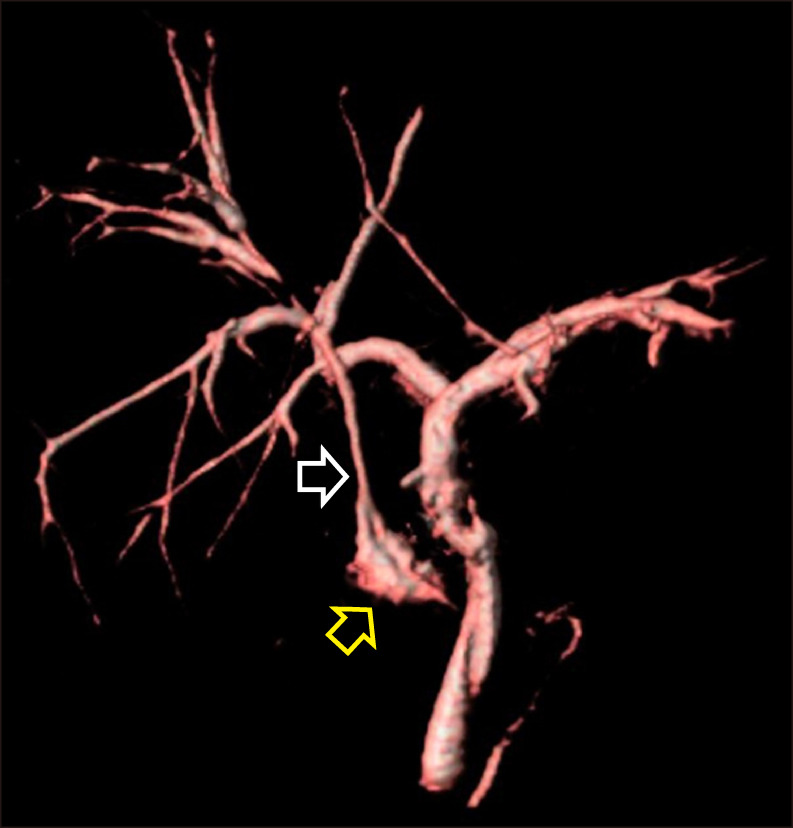
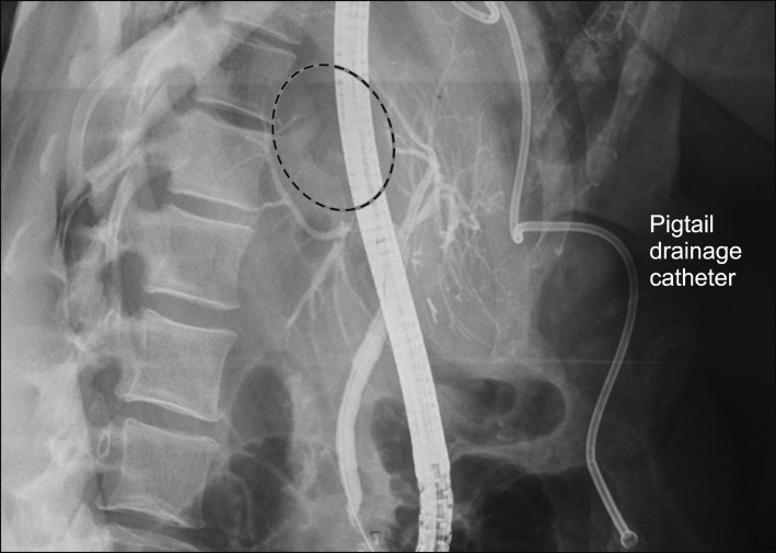
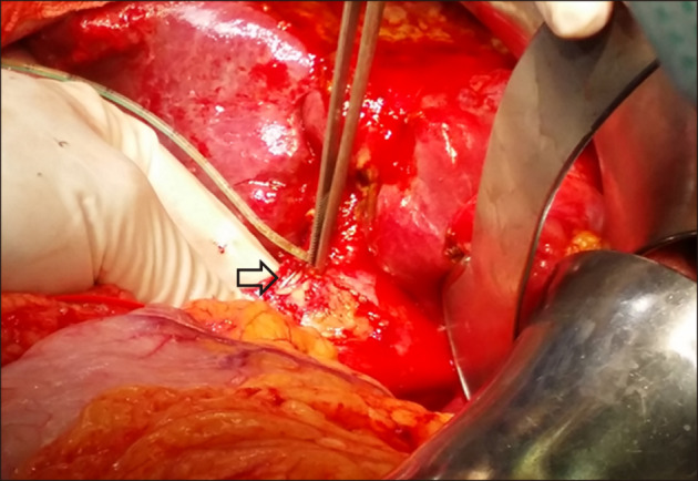
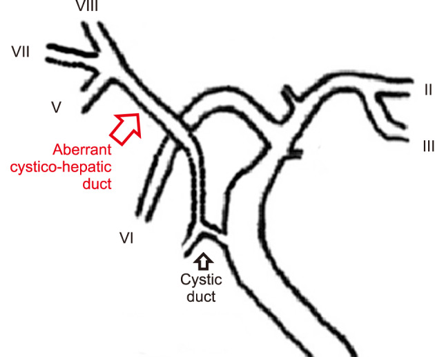
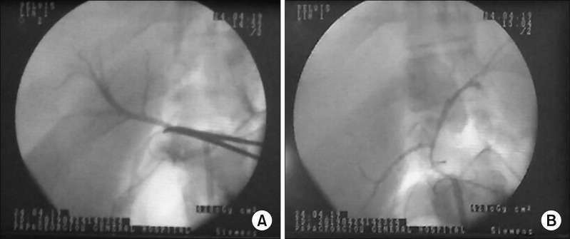
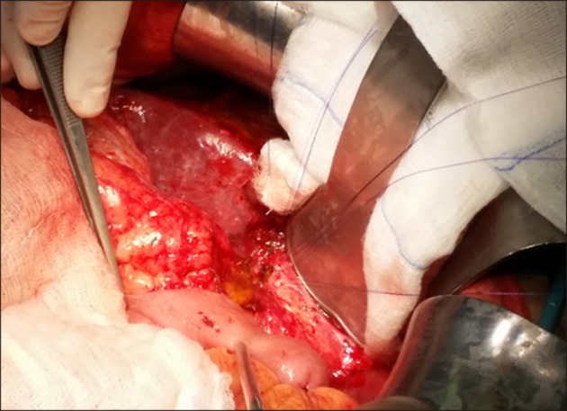
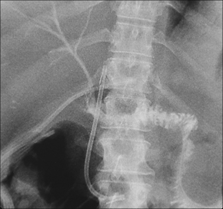
A typical bile duct branching patterns represent one of the major causes of bile duct injury (BDI) during laparoscopic cholecystectomy (LC). The most common classified variations of bile duct branching, involve the right posterior sectoral duct (RPSD) and its joining with the right anterior or left hepatic duct. Variant bile duct anatomy can rarely be extremely complex and unclassified. This report describes an extremely rare case of an isolated injury to an aberrant right hepatic duct formed by the joining of ducts from segments V, VII, and VIII draining into the cystic duct (cysticohepatic duct) during LC, associated with an inferior RPSD opening to left hepatic duct. Detailed evaluation of both endoscopic and magnetic cholangiograms established the diagnosis. Bile duct injury was subsequently managed surgically by a demanding Roux-en-Y hepaticojejunostomy. This extremely rare case aims to serve as a useful reminder of the consistent inconsistency of biliary anatomy, alerting surgeons to beware of variant bile duct branching patterns during open or LC that constitute a dreadful pitfall for severe and life-threatening bile duct injuries.
典型的胆管分支模式是腹腔镜胆囊切除术(LC)期间胆管损伤(BDI)的主要原因之一。胆管分支最常见的分类变异涉及右后叶胆管(RPSD)及其与右前叶或左肝管的汇合。变异的胆管解剖结构可能极其复杂且难以分类。本报告描述了一例极其罕见的病例,在LC期间,由V、VII和VIII段胆管汇合形成的异常右肝管汇入胆囊管(胆囊肝管),导致孤立性损伤,并伴有低位RPSD开口于左肝管。通过内镜和磁共振胆管造影进行详细评估后确诊。随后通过要求较高的Roux-en-Y肝空肠吻合术对胆管损伤进行了手术处理。这一极其罕见的病例旨在提醒人们注意胆管解剖结构始终存在的不一致性,提醒外科医生在开腹手术或LC期间要警惕变异的胆管分支模式,因为这可能会导致严重且危及生命的胆管损伤。