Department of Biology, Johns Hopkins University, Baltimore, Maryland, United States of America.
Department of Biophysics, Johns Hopkins University, Baltimore, Maryland, United States of America.
PLoS Comput Biol. 2020 Mar 9;16(3):e1007691. doi: 10.1371/journal.pcbi.1007691. eCollection 2020 Mar.
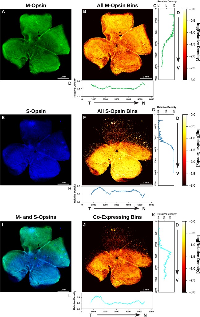
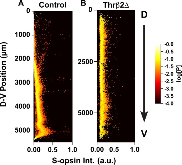
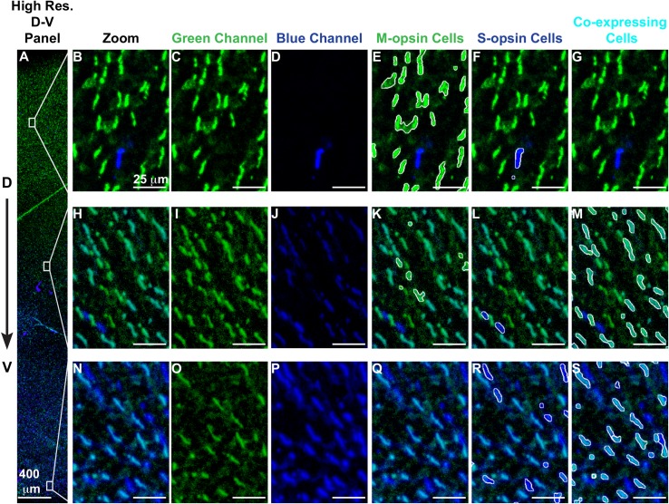
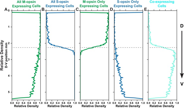
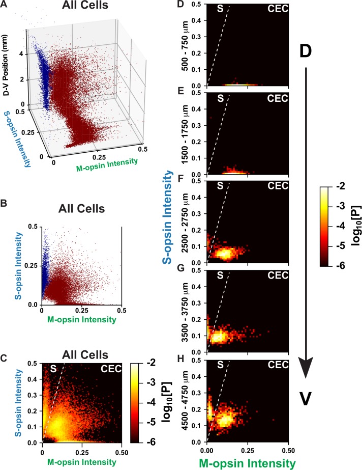
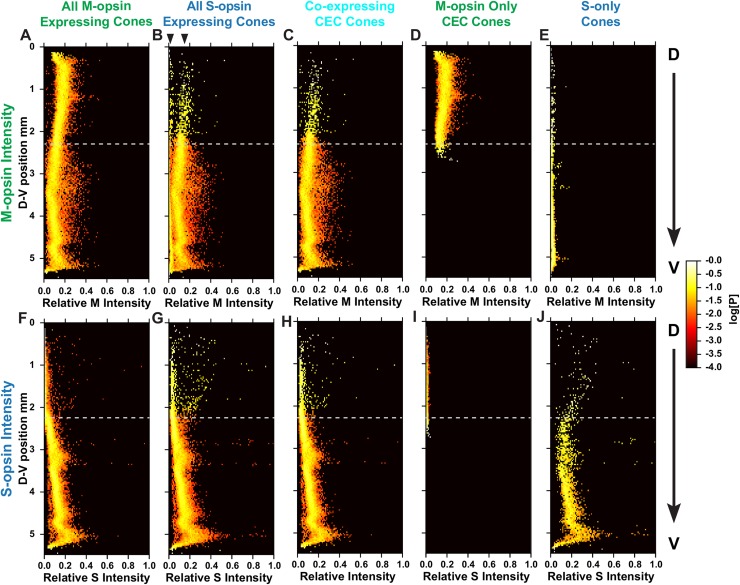
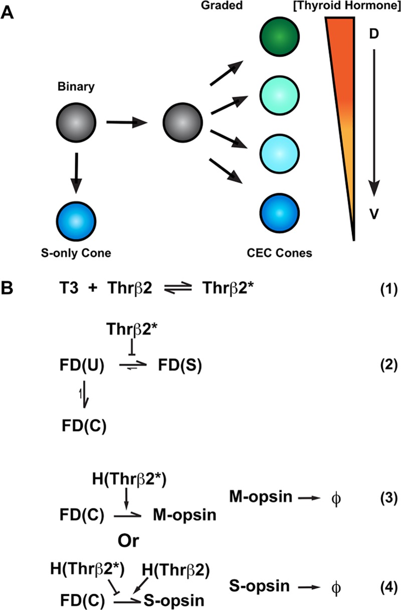
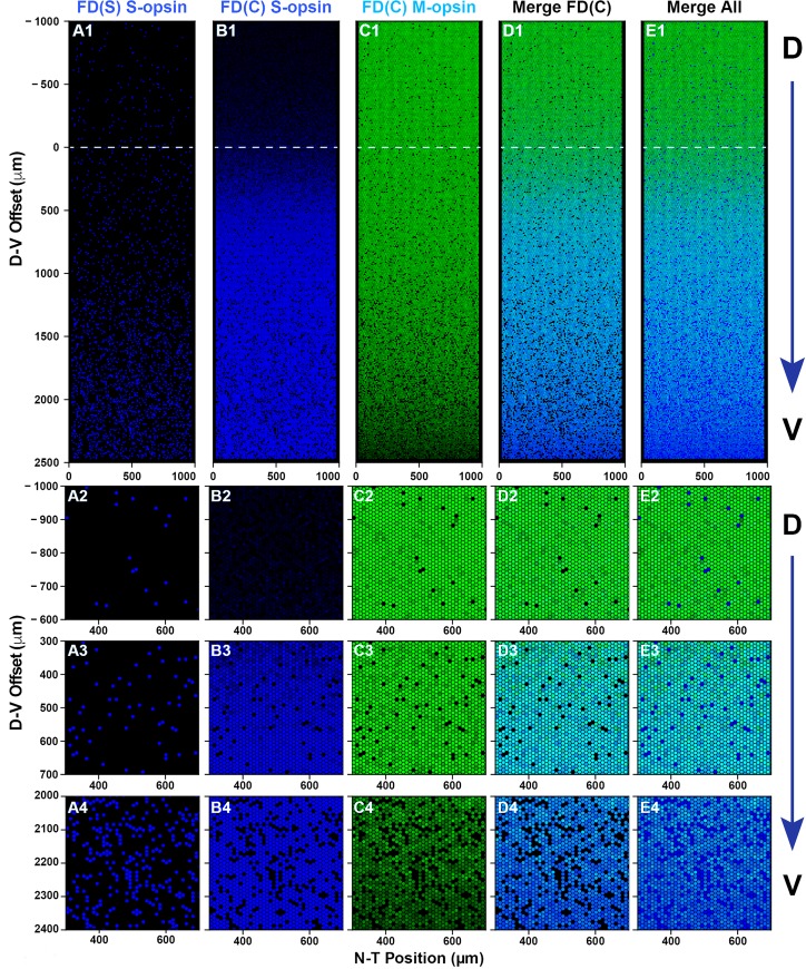
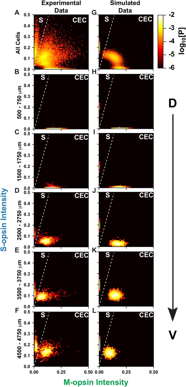
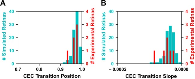
Nervous systems are incredibly diverse, with myriad neuronal subtypes defined by gene expression. How binary and graded fate characteristics are patterned across tissues is poorly understood. Expression of opsin photopigments in the cone photoreceptors of the mouse retina provides an excellent model to address this question. Individual cones express S-opsin only, M-opsin only, or both S-opsin and M-opsin. These cell populations are patterned along the dorsal-ventral axis, with greater M-opsin expression in the dorsal region and greater S-opsin expression in the ventral region. Thyroid hormone signaling plays a critical role in activating M-opsin and repressing S-opsin. Here, we developed an image analysis approach to identify individual cone cells and evaluate their opsin expression from immunofluorescence imaging tiles spanning roughly 6 mm along the D-V axis of the mouse retina. From analyzing the opsin expression of ~250,000 cells, we found that cones make a binary decision between S-opsin only and co-expression competent fates. Co-expression competent cells express graded levels of S- and M-opsins, depending nonlinearly on their position in the dorsal-ventral axis. M- and S-opsin expression display differential, inverse patterns. Using these single-cell data, we developed a quantitative, probabilistic model of cone cell decisions in the retinal tissue based on thyroid hormone signaling activity. The model recovers the probability distribution for cone fate patterning in the mouse retina and describes a minimal set of interactions that are necessary to reproduce the observed cell fates. Our study provides a paradigm describing how differential responses to regulatory inputs generate complex patterns of binary and graded cell fates.
神经系统具有惊人的多样性,其神经元亚型可通过基因表达来定义。人们对组织中二元和渐变命运特征的模式形成了解甚少。在小鼠视网膜的视锥光感受器中表达视蛋白光色素,为解决这个问题提供了一个极好的模型。单个视锥仅表达 S-视蛋白、M-视蛋白或 S-视蛋白和 M-视蛋白都表达。这些细胞群体沿着背腹轴模式化,在背侧区域表达更多的 M-视蛋白,在腹侧区域表达更多的 S-视蛋白。甲状腺激素信号在激活 M-视蛋白和抑制 S-视蛋白方面起着至关重要的作用。在这里,我们开发了一种图像分析方法,从跨越小鼠视网膜背腹轴约 6 毫米的免疫荧光成像平铺中识别单个视锥细胞并评估其视蛋白表达。通过分析大约 250,000 个细胞的视蛋白表达,我们发现视锥细胞在 S-视蛋白仅表达和共表达能力命运之间做出了二元决定。共表达能力的细胞表达 S-和 M-视蛋白的分级水平,这取决于它们在背腹轴上的位置呈非线性关系。M-和 S-视蛋白表达显示出不同的、相反的模式。使用这些单细胞数据,我们基于甲状腺激素信号活性,为视网膜组织中的视锥细胞决策开发了一个定量的、概率模型。该模型恢复了小鼠视网膜中视锥命运模式形成的概率分布,并描述了一组必要的最小相互作用,以重现观察到的细胞命运。我们的研究提供了一个描述如何对调节输入产生复杂的二元和渐变细胞命运模式的范例。