Department of Diagnostic Radiology, "San Gerardo" Hospital, Via Pergolesi 33, 20900, Monza, MB, Italy; School of Medicine, University of Milano-Bicocca, Via Cadore 48, 20900, Monza, MB, Italy.
Department of Diagnostic Radiology, "San Gerardo" Hospital, Via Pergolesi 33, 20900, Monza, MB, Italy; School of Medicine, University of Milano-Bicocca, Via Cadore 48, 20900, Monza, MB, Italy.
Respir Med. 2020 Aug-Sep;170:106036. doi: 10.1016/j.rmed.2020.106036. Epub 2020 May 22.
To evaluate the imaging features of routine admission chest X-ray in patients referred for novel Coronavirus 2019 infection.
All patients referred to the emergency departments, RT-PCR positive for SARS-CoV-2 infection were evaluated. Demographic and clinical data were recorded. Two radiologists (8 and 15 years of experience) reviewed all the X-ray images and evaluated the following findings: interstitial opacities, alveolar opacities (AO), AO associated with consolidation, consolidation and/or pleural effusion. We stratified patients in groups according to the time interval between symptoms onset (cut-off 5 days) and X-ray imaging and according to age (cut-off 60 years old). Computed tomography was performed in case of a discrepancy between clinical symptoms, laboratory and X-ray findings, and/or suspicion of complications.
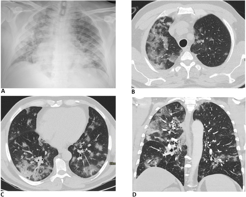
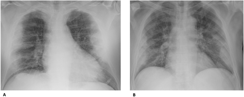
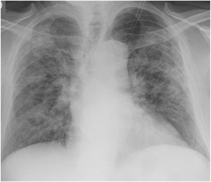
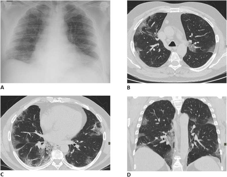
A total of 468 patients were tested positive for SARS-CoV-2. Lung lesions primarily manifested as interstitial opacities (71.7%) and AO opacities (60.5%), more frequently bilateral (64.5%) and with a peripheral predominance (62.5%). Patients admitted to the emergency radiology department after 5 days from symptoms onset, more frequently had interstitial and AO opacities, in comparison to those admitted within 5 days, and lung lesions were more frequently bilateral and peripheral. Older patients more frequently presented interstitial and AO opacities in comparison to younger ones. Sixty-eight patients underwent CT that principally showed the presence of ground-glass opacities and consolidations.
The most common X-ray pattern is multifocal and peripheral, associated with interstitial and alveolar opacities. Chest X-ray, compared to CT, can be considered a reliable diagnostic tool, especially in the Emergency setting.
评估新型冠状病毒 2019 感染患者常规入院胸部 X 线的影像学特征。
对因新型冠状病毒 2019 感染而被转至急诊部、实时逆转录聚合酶链反应(RT-PCR)检测阳性的所有患者进行评估。记录人口统计学和临床数据。两位放射科医生(8 年和 15 年经验)分别回顾所有 X 射线图像,并评估以下发现:间质混浊、肺泡混浊(AO)、与实变相关的 AO、实变和/或胸腔积液。我们根据症状发作(截止日期为 5 天)和 X 射线成像之间的时间间隔以及根据年龄(截止日期为 60 岁)将患者分为组。如果临床症状、实验室和 X 射线结果之间存在差异,以及/或怀疑有并发症,则进行计算机断层扫描(CT)检查。
共有 468 例患者被检测出新型冠状病毒 2019 呈阳性。肺部病变主要表现为间质混浊(71.7%)和 AO 混浊(60.5%),更常为双侧(64.5%),且具有外周优势(62.5%)。与症状发作后 5 天内入院的患者相比,症状发作后 5 天内入院的患者更常出现间质和 AO 混浊,且肺部病变更常为双侧和外周。与年轻患者相比,老年患者更常出现间质混浊和 AO 混浊。68 例患者进行了 CT 检查,主要显示磨玻璃混浊和实变的存在。
最常见的 X 射线表现是多灶性和外周性,伴有间质和肺泡混浊。与 CT 相比,X 射线可被视为一种可靠的诊断工具,尤其是在急诊环境中。