Department of Radiology, Jiangxi Provincial People's Hospital, Nanchang, China.
Department of Medical Cosmetology, Jiangxi Provincial People's Hospital, Nanchang, China.
Korean J Radiol. 2020 Apr;21(4):501-504. doi: 10.3348/kjr.2020.0112. Epub 2020 Feb 26.
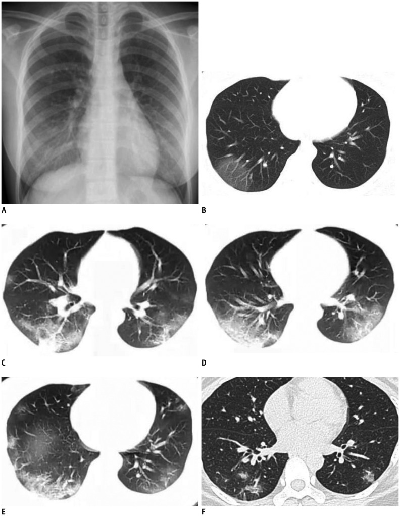
From December 2019, Coronavirus disease 2019 (COVID-19) pneumonia (formerly known as the 2019 novel Coronavirus [2019-nCoV]) broke out in Wuhan, China. In this study, we present serial CT findings in a 40-year-old female patient with COVID-19 pneumonia who presented with the symptoms of fever, chest tightness, and fatigue. She was diagnosed with COVID-19 infection confirmed by real-time reverse-transcriptase-polymerase chain reaction. CT showed rapidly progressing peripheral consolidations and ground-glass opacities in both lungs. After treatment, the lesions were shown to be almost absorbed leaving the fibrous lesions.
从 2019 年 12 月开始,中国武汉爆发了 2019 年冠状病毒病(COVID-19)肺炎(以前称为 2019 年新型冠状病毒[2019-nCoV])。在本研究中,我们呈现了一位 40 岁女性 COVID-19 肺炎患者的连续 CT 表现,该患者有发热、胸闷和乏力的症状。她被实时逆转录-聚合酶链反应诊断为 COVID-19 感染。CT 显示双肺外周迅速进展的实变和磨玻璃影。经过治疗,病变几乎被吸收,留下纤维病变。