Wang Man-Man, Zhang Min, Feng Ya-Shuo, Xing Ying, Tan Zi-Xuan, Li Wen-Bin, Dong Fang, Zhang Feng
Department of Rehabilitation Medicine, The Third Hospital of Hebei Medical University, Shijiazhuang, China.
Department of Pathophysiology, Hebei Medical University, Shijiazhuang, China.
Front Cell Neurosci. 2020 May 15;14:134. doi: 10.3389/fncel.2020.00134. eCollection 2020.
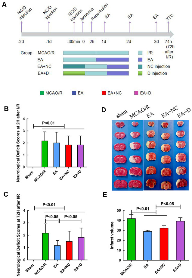
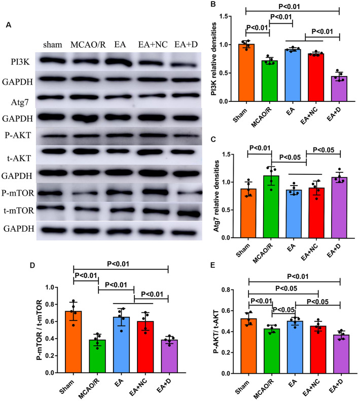
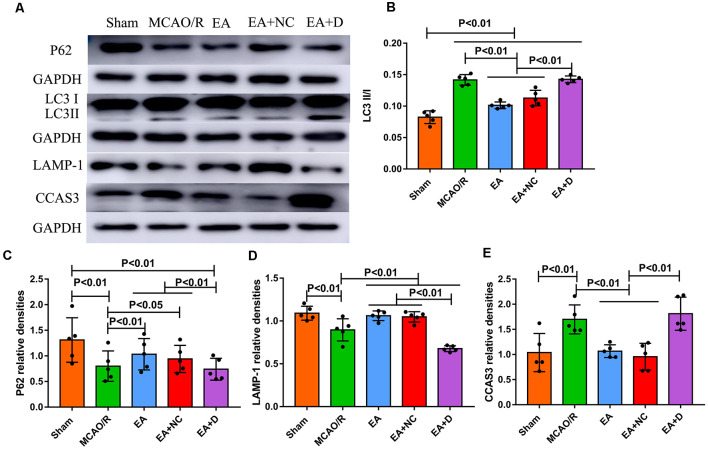
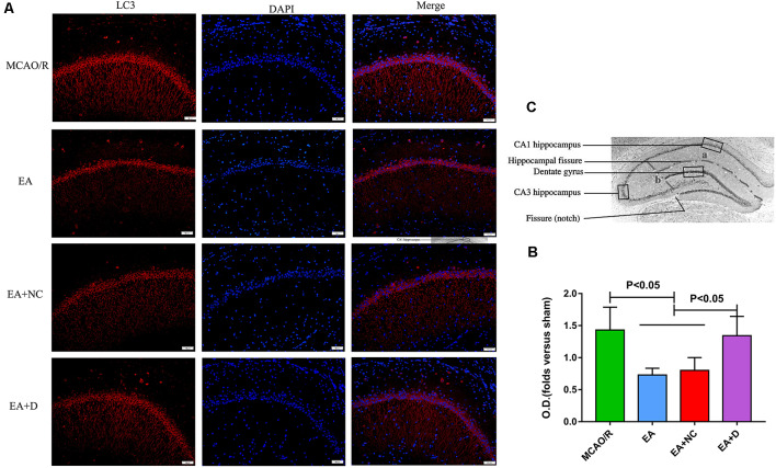
Electroacupuncture (EA) is a safe and effective therapy for ischemic stroke in both clinical and laboratory settings. However, the underlying mechanism behind EA treatment for stroke remains unclear. Here, we aimed to evaluate whether EA treatment at the acupoints of Zusanli (ST36) and Quchi (LI11) exerted a neuroprotective effect on ischemic stroke rats by modulating autophagy and apoptosis the PI3K/AKT/mTOR signaling pathway. EA was performed at 24 h following brain ischemia/reperfusion (I/R) for 30 min per day for 3 days. Our results indicated that EA treatment significantly decreased neurological deficits and cerebral infarct volume in ischemic stroke rats. Also, EA intervention markedly reduced neuronal apoptosis by suppressing the activation of cleaved caspase-3 (CCAS3) at 72 h following I/R, as shown by a Western blot analysis. Furthermore, EA treatment after ischemic stroke suppressed the ischemia activated expression level of LC3II/I and Atg7 and increased the ischemia inhibited expression level of PI3K, phosphorylation of mTOR, phosphorylation of AKT, P62 and LAMP1, hence mediating the autophagy level of the neurocyte, which was reversed by the PI3K inhibitor Dactolisib. In summary, our results indicate that the protective effects of EA treatment at points of Quchi (LI11) and Zusanli (ST36) in rats following cerebral I/R injury was associated with the inhibition of neuronal apoptosis and autophagy activating the PI3K/AKT/mTOR signaling pathway.
在临床和实验室环境中,电针(EA)都是治疗缺血性中风的一种安全有效的疗法。然而,EA治疗中风的潜在机制仍不清楚。在此,我们旨在评估在足三里(ST36)和曲池(LI11)穴位进行EA治疗是否通过调节自噬和凋亡以及PI3K/AKT/mTOR信号通路,对缺血性中风大鼠发挥神经保护作用。在脑缺血/再灌注(I/R)后24小时开始进行EA治疗,每天30分钟,持续3天。我们的结果表明,EA治疗显著降低了缺血性中风大鼠的神经功能缺损和脑梗死体积。此外,如蛋白质免疫印迹分析所示,EA干预在I/R后72小时通过抑制裂解的半胱天冬酶-3(CCAS3)的激活,显著减少了神经元凋亡。此外,缺血性中风后的EA治疗抑制了缺血激活的LC3II/I和Atg7的表达水平,并增加了缺血抑制的PI3K、mTOR磷酸化、AKT磷酸化、P62和LAMP1的表达水平,从而调节神经细胞的自噬水平,而PI3K抑制剂哌立福新可逆转这种调节。总之,我们的结果表明,在大鼠脑I/R损伤后,在曲池(LI11)和足三里(ST36)穴位进行EA治疗的保护作用与抑制神经元凋亡和自噬以及激活PI3K/AKT/mTOR信号通路有关。