Shen Boyuan, Chen Xiao, Shen Kui, Xiong Hao, Wei Fei
Beijing Key Laboratory of Green Chemical Reaction Engineering and Technology, Department of Chemical Engineering, Tsinghua University, Beijing, 100084, China.
School of Chemistry and Chemical Engineering, South China University of Technology, Guangzhou, 510640, China.
Nat Commun. 2020 Jun 1;11(1):2692. doi: 10.1038/s41467-020-16531-y.
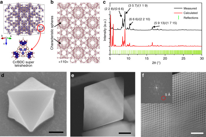
Porous metal-organic frameworks (MOFs) have shown wide applications in catalysis, gas storage and separation due to their highly tunable porosity, connectivity and local structures. However, the electron-beam sensitivity of MOFs makes it difficult to achieve the atomic imaging of their bulk and local structures under (scanning) transmission electron microscopy ((S)TEM) to study their structure-property relations. Here, we report the low-dose imaging of a beam-sensitive MOF, MIL-101, under a Cs-corrected STEM based on the integrated differential phase contrast (iDPC) technique. The images resolve the coordination of Cr nodes and organic linkers inside the frameworks with an information transfer of ~1.8Å. The local structures in MIL-101 are also revealed under iDPC-STEM, including the surfaces, interfaces and defects. These results provide an extensible method to image various beam-sensitive materials with ultrahigh resolution, and unravel the whole framework architectures for further defect and surface engineering of MOFs towards tailored functions.
多孔金属有机框架材料(MOFs)因其高度可调的孔隙率、连接性和局部结构,在催化、气体储存和分离等领域展现出广泛应用。然而,MOFs对电子束的敏感性使得在(扫描)透射电子显微镜((S)TEM)下难以实现其整体和局部结构的原子成像,从而难以研究其结构-性能关系。在此,我们基于积分差分相衬(iDPC)技术,报道了在Cs校正扫描透射电子显微镜(STEM)下对束敏感型MOF材料MIL-101进行的低剂量成像。这些图像以约1.8Å的信息传递分辨率解析了框架内Cr节点与有机连接体的配位情况。在iDPC-STEM下还揭示了MIL-101的局部结构,包括表面、界面和缺陷。这些结果提供了一种可扩展的方法,用于以超高分辨率对各种束敏感材料进行成像,并揭示整个框架结构,以便对MOFs进行进一步的缺陷和表面工程设计,实现定制功能。