• 文献检索
  • 文档翻译
  • 深度研究
  • 学术资讯
  • Suppr Zotero 插件Zotero 插件
  • 邀请有礼
  • 套餐&价格
  • 历史记录
应用&插件
Suppr Zotero 插件Zotero 插件浏览器插件Mac 客户端Windows 客户端微信小程序
定价
高级版会员购买积分包购买API积分包
服务
文献检索文档翻译深度研究API 文档MCP 服务
关于我们
关于 Suppr公司介绍联系我们用户协议隐私条款
关注我们

Suppr 超能文献

核心技术专利:CN118964589B侵权必究
粤ICP备2023148730 号-1Suppr @ 2026

文献检索

告别复杂PubMed语法,用中文像聊天一样搜索,搜遍4000万医学文献。AI智能推荐,让科研检索更轻松。

立即免费搜索

文件翻译

保留排版,准确专业,支持PDF/Word/PPT等文件格式,支持 12+语言互译。

免费翻译文档

深度研究

AI帮你快速写综述,25分钟生成高质量综述,智能提取关键信息,辅助科研写作。

立即免费体验

iDPC-STEM 的发展及其在电子束敏感材料中的应用。

The Development of iDPC-STEM and Its Application in Electron Beam Sensitive Materials.

机构信息

Beijing Key Laboratory of Microstructure and Properties of Solids, Faculty of Materials and Manufacturing, Beijing University of Technique, Beijing 100124, China.

出版信息

Molecules. 2022 Jun 14;27(12):3829. doi: 10.3390/molecules27123829.

DOI:10.3390/molecules27123829
PMID:35744947
原文链接:https://pmc.ncbi.nlm.nih.gov/articles/PMC9231126/
Abstract

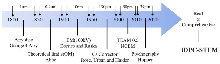
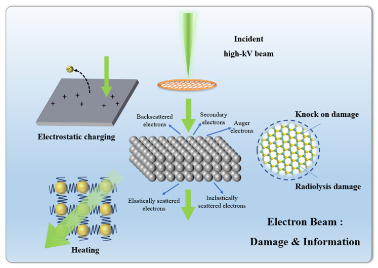
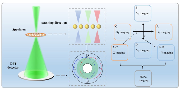
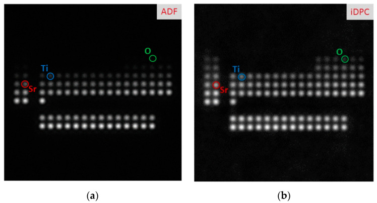
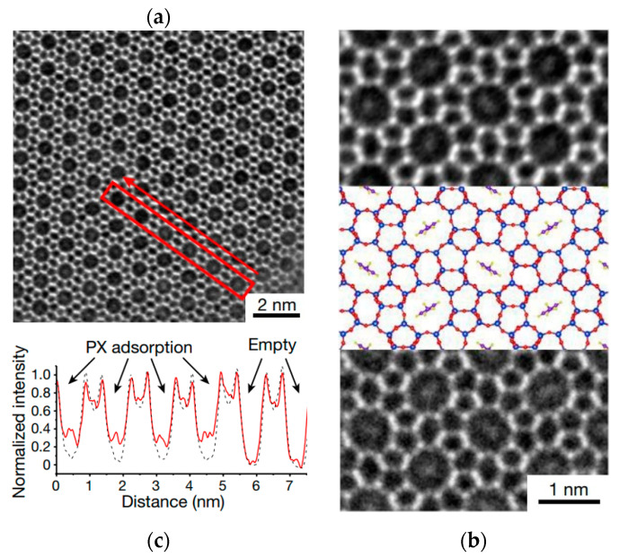
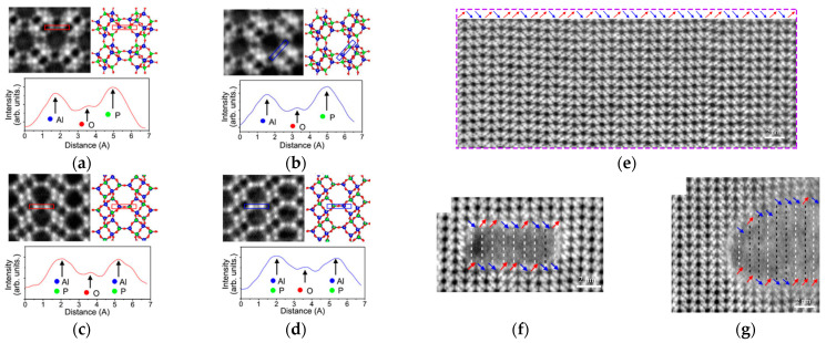
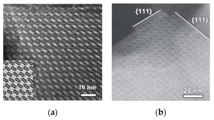
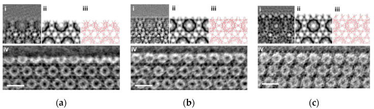
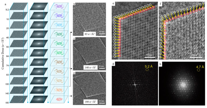
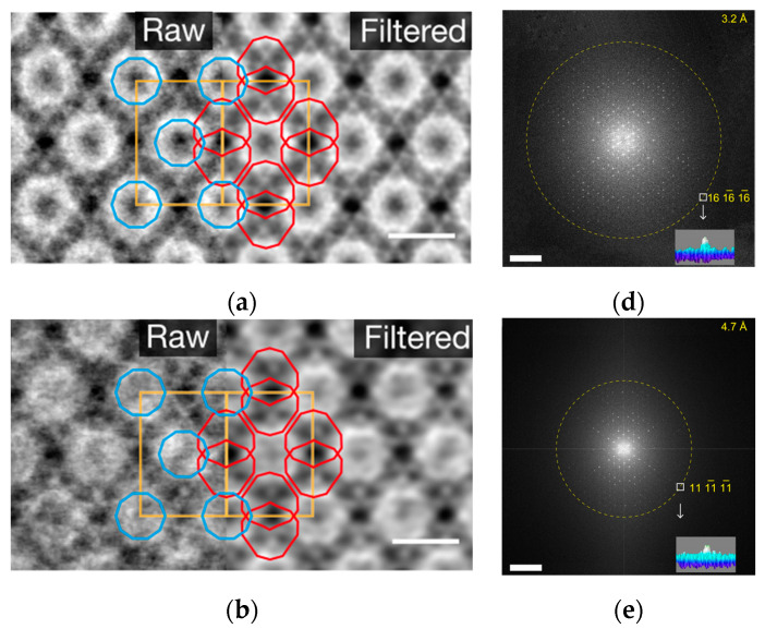
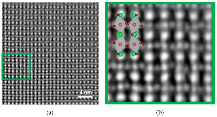
The main aspects of material research: material synthesis, material structure, and material properties, are interrelated. Acquiring atomic structure information of electron beam sensitive materials by electron microscope, such as porous zeolites, organic-inorganic hybrid perovskites, metal-organic frameworks, is an important and challenging task. The difficulties in characterization of the structures will inevitably limit the optimization of their synthesis methods and further improve their performance. The emergence of integrated differential phase contrast scanning transmission electron microscopy (iDPC-STEM), a STEM characterization technique capable of obtaining images with high signal-to-noise ratio under lower doses, has made great breakthroughs in the atomic structure characterization of these materials. This article reviews the developments and applications of iDPC-STEM in electron beam sensitive materials, and provides an outlook on its capabilities and development.

摘要

材料研究的主要方面

材料合成、材料结构和材料性能,是相互关联的。通过电子显微镜获取电子束敏感材料(如多孔沸石、有机-无机杂化钙钛矿、金属有机骨架)的原子结构信息,是一项重要且具有挑战性的任务。结构特征的困难必然会限制其合成方法的优化,并进一步提高其性能。集成微分相衬扫描透射电子显微镜(iDPC-STEM)的出现,是一种能够在较低剂量下获得高信噪比图像的 STEM 表征技术,在这些材料的原子结构表征方面取得了重大突破。本文综述了 iDPC-STEM 在电子束敏感材料中的发展和应用,并对其能力和发展前景进行了展望。

https://cdn.ncbi.nlm.nih.gov/pmc/blobs/1049/9231126/c6c79eab32af/molecules-27-03829-g022.jpg
https://cdn.ncbi.nlm.nih.gov/pmc/blobs/1049/9231126/59b3d6bee0d0/molecules-27-03829-g001.jpg
https://cdn.ncbi.nlm.nih.gov/pmc/blobs/1049/9231126/74d4b562f68a/molecules-27-03829-g002.jpg
https://cdn.ncbi.nlm.nih.gov/pmc/blobs/1049/9231126/a9a6e05f3393/molecules-27-03829-g003.jpg
https://cdn.ncbi.nlm.nih.gov/pmc/blobs/1049/9231126/22d52518c065/molecules-27-03829-g004.jpg
https://cdn.ncbi.nlm.nih.gov/pmc/blobs/1049/9231126/a652a750b2e3/molecules-27-03829-g005.jpg
https://cdn.ncbi.nlm.nih.gov/pmc/blobs/1049/9231126/dff9f679222f/molecules-27-03829-g006.jpg
https://cdn.ncbi.nlm.nih.gov/pmc/blobs/1049/9231126/b2ebd77fb6bf/molecules-27-03829-g007.jpg
https://cdn.ncbi.nlm.nih.gov/pmc/blobs/1049/9231126/337dbfac1262/molecules-27-03829-g008.jpg
https://cdn.ncbi.nlm.nih.gov/pmc/blobs/1049/9231126/943296b83b7b/molecules-27-03829-g009a.jpg
https://cdn.ncbi.nlm.nih.gov/pmc/blobs/1049/9231126/64928d290b73/molecules-27-03829-g010.jpg
https://cdn.ncbi.nlm.nih.gov/pmc/blobs/1049/9231126/f67d17c0885d/molecules-27-03829-g011.jpg
https://cdn.ncbi.nlm.nih.gov/pmc/blobs/1049/9231126/09a0e4133238/molecules-27-03829-g012.jpg
https://cdn.ncbi.nlm.nih.gov/pmc/blobs/1049/9231126/2b7408972955/molecules-27-03829-g013.jpg
https://cdn.ncbi.nlm.nih.gov/pmc/blobs/1049/9231126/1e00a0eb7853/molecules-27-03829-g014.jpg
https://cdn.ncbi.nlm.nih.gov/pmc/blobs/1049/9231126/1817d096f68e/molecules-27-03829-g015.jpg
https://cdn.ncbi.nlm.nih.gov/pmc/blobs/1049/9231126/f4567217837c/molecules-27-03829-g016a.jpg
https://cdn.ncbi.nlm.nih.gov/pmc/blobs/1049/9231126/76b6ae6e37fa/molecules-27-03829-g017.jpg
https://cdn.ncbi.nlm.nih.gov/pmc/blobs/1049/9231126/f5147ba1de5d/molecules-27-03829-g018.jpg
https://cdn.ncbi.nlm.nih.gov/pmc/blobs/1049/9231126/a087a68f5702/molecules-27-03829-g019a.jpg
https://cdn.ncbi.nlm.nih.gov/pmc/blobs/1049/9231126/8328a8d46918/molecules-27-03829-g020.jpg
https://cdn.ncbi.nlm.nih.gov/pmc/blobs/1049/9231126/87fb391d3327/molecules-27-03829-g021.jpg
https://cdn.ncbi.nlm.nih.gov/pmc/blobs/1049/9231126/c6c79eab32af/molecules-27-03829-g022.jpg

相似文献

1
The Development of iDPC-STEM and Its Application in Electron Beam Sensitive Materials.iDPC-STEM 的发展及其在电子束敏感材料中的应用。
Molecules. 2022 Jun 14;27(12):3829. doi: 10.3390/molecules27123829.
2
Imaging biological samples by integrated differential phase contrast (iDPC) STEM technique.通过集成差分相衬(iDPC)扫描透射电子显微镜技术对生物样品进行成像。
J Struct Biol. 2022 Mar;214(1):107837. doi: 10.1016/j.jsb.2022.107837. Epub 2022 Jan 31.
3
Atomic Spatial and Temporal Imaging of Local Structures and Light Elements inside Zeolite Frameworks.沸石骨架中局部结构和轻元素的原子空间和时空成像。
Adv Mater. 2020 Jan;32(4):e1906103. doi: 10.1002/adma.201906103. Epub 2019 Nov 29.
4
Optimizing experimental parameters of integrated differential phase contrast (iDPC) for atomic resolution imaging.优化集成差分相衬(iDPC)的实验参数以实现原子分辨率成像。
Ultramicroscopy. 2023 Apr;246:113686. doi: 10.1016/j.ultramic.2023.113686. Epub 2023 Jan 18.
5
Imaging the node-linker coordination in the bulk and local structures of metal-organic frameworks.成像金属有机框架的整体和局部结构中的节点-连接体配位情况。
Nat Commun. 2020 Jun 1;11(1):2692. doi: 10.1038/s41467-020-16531-y.
6
Imaging Beam-Sensitive Materials by Electron Microscopy.通过电子显微镜对光束敏感材料进行成像。
Adv Mater. 2020 Apr;32(16):e1907619. doi: 10.1002/adma.201907619. Epub 2020 Feb 28.
7
Real-Space Imaging of the Node-Linker Coordination on the Interfaces between Self-Assembled Metal-Organic Frameworks.自组装金属有机框架界面上节点-连接体配位的实空间成像
Nano Lett. 2022 Dec 28;22(24):9928-9934. doi: 10.1021/acs.nanolett.2c03375. Epub 2022 Dec 13.
8
High-resolution imaging of organic pharmaceutical crystals by transmission electron microscopy and scanning moiré fringes.利用透射电子显微镜和扫描云纹条纹对有机药物晶体进行高分辨率成像。
J Microsc. 2020 Sep;279(3):197-206. doi: 10.1111/jmi.12866. Epub 2020 Feb 18.
9
Tutorial: structural characterization of isolated metal atoms and subnanometric metal clusters in zeolites.教程:沸石中孤立金属原子和亚纳米金属团簇的结构特征。
Nat Protoc. 2021 Apr;16(4):1871-1906. doi: 10.1038/s41596-020-0366-9. Epub 2020 Sep 4.
10
Real-Space Imaging of the Molecular Changes in Metal-Organic Frameworks under Electron Irradiation.电子辐照下金属有机框架中分子变化的实空间成像
ACS Nano. 2023 Mar 14;17(5):4740-4747. doi: 10.1021/acsnano.2c11110. Epub 2023 Feb 22.

引用本文的文献

1
Simulation Study of Low-Dose 4D-STEM Phase Contrast Techniques at the Nanoscale in SEM.扫描电子显微镜中纳米尺度低剂量4D-STEM相衬技术的模拟研究
Nanomaterials (Basel). 2025 Jan 4;15(1):70. doi: 10.3390/nano15010070.
2
Low-cost electron detector for scanning electron microscope.用于扫描电子显微镜的低成本电子探测器。
HardwareX. 2023 Mar 10;14:e00413. doi: 10.1016/j.ohx.2023.e00413. eCollection 2023 Jun.

本文引用的文献

1
Bulk and local structures of metal-organic frameworks unravelled by high-resolution electron microscopy.通过高分辨率电子显微镜揭示金属有机框架的整体和局部结构。
Commun Chem. 2020 Aug 6;3(1):99. doi: 10.1038/s42004-020-00361-6.
2
Molecular-Level Insight into Correlation between Surface Defects and Stability of Methylammonium Lead Halide Perovskite Under Controlled Humidity.湿度可控条件下甲基铵卤化铅钙钛矿表面缺陷与稳定性之间相关性的分子水平洞察
Small Methods. 2021 Feb;5(2):e2000834. doi: 10.1002/smtd.202000834. Epub 2020 Nov 3.
3
Cryo-EM structures of atomic surfaces and host-guest chemistry in metal-organic frameworks.
金属有机框架中原子表面的冷冻电镜结构及主客体化学
Matter. 2019 Aug 7;1(2):428-438. doi: 10.1016/j.matt.2019.06.001. Epub 2020 Mar 24.
4
Electron ptychography achieves atomic-resolution limits set by lattice vibrations.电子相衬层析成像达到了由晶格振动设定的原子分辨率极限。
Science. 2021 May 21;372(6544):826-831. doi: 10.1126/science.abg2533.
5
A single-molecule van der Waals compass.单分子范德瓦尔斯指南针。
Nature. 2021 Apr;592(7855):541-544. doi: 10.1038/s41586-021-03429-y. Epub 2021 Apr 21.
6
Resolving atomic SAPO-34/18 intergrowth architectures for methanol conversion by identifying light atoms and bonds.通过识别轻原子和键来解析用于甲醇转化的原子级SAPO-34/18共生结构。
Nat Commun. 2021 Apr 13;12(1):2212. doi: 10.1038/s41467-021-22438-z.
7
Single-Crystalline Ultrathin 2D Porous Nanosheets of Chiral Metal-Organic Frameworks.手性金属-有机框架的单晶超薄 2D 多孔纳米片
J Am Chem Soc. 2021 Mar 10;143(9):3509-3518. doi: 10.1021/jacs.0c13005. Epub 2021 Feb 23.
8
Filling metal-organic framework mesopores with TiO for CO photoreduction.用 TiO 填充金属-有机骨架介孔以进行 CO 光还原。
Nature. 2020 Oct;586(7830):549-554. doi: 10.1038/s41586-020-2738-2. Epub 2020 Sep 9.
9
Structures and Structural Evolution of Sublayer Surfaces of Metal-Organic Frameworks.金属有机框架材料亚层表面的结构与结构演变
Angew Chem Int Ed Engl. 2020 Nov 23;59(48):21419-21424. doi: 10.1002/anie.202008100. Epub 2020 Sep 23.
10
Direct Imaging of Correlated Defect Nanodomains in a Metal-Organic Framework.金属有机框架中相关缺陷纳米域的直接成像
J Am Chem Soc. 2020 Jul 29;142(30):13081-13089. doi: 10.1021/jacs.0c04468. Epub 2020 Jul 16.