Karagul Meryem Ilkay, Aktas Savas, Yilmaz Sakir Necat, Yetkin Derya, Celikcan Havva Didem, Cevik Ozge Selin
Department of Histology and Embryology, Faculty of Medicine, Mersin University, Mersin, Turkey.
Advanced Technology of Education, Research and Application Center, Mersin University, Mersin, Turkey.
EXCLI J. 2020 May 4;19:532-546. doi: 10.17179/excli2019-1834. eCollection 2020.
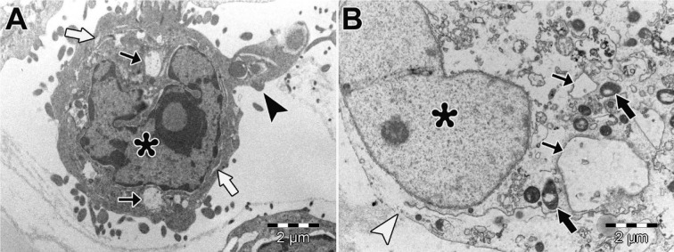
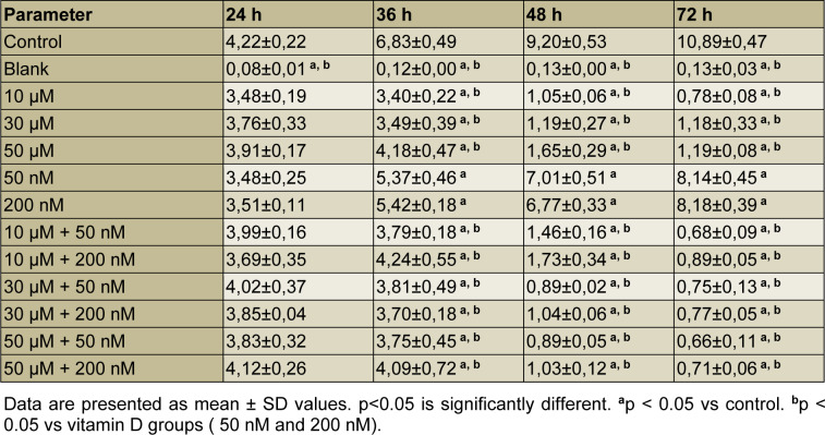
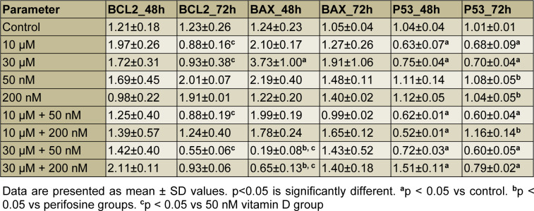
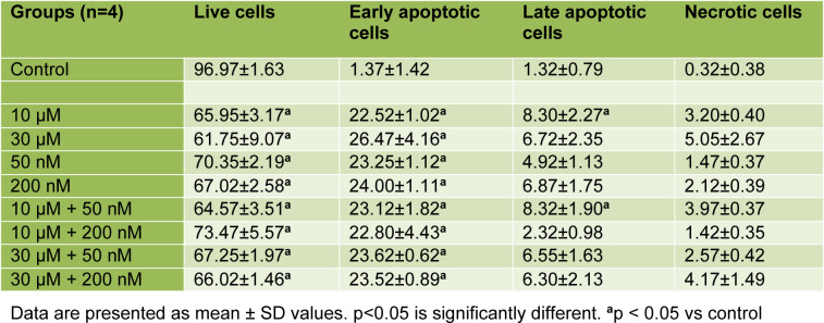
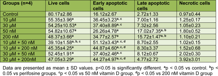
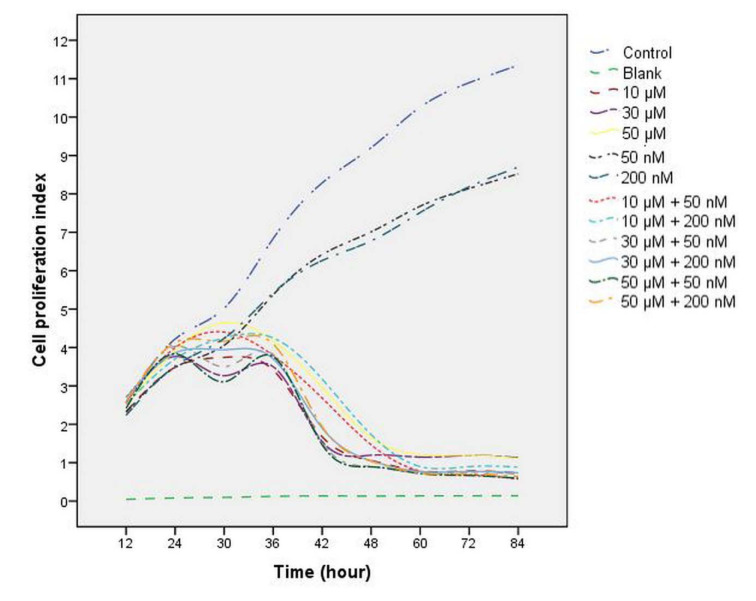
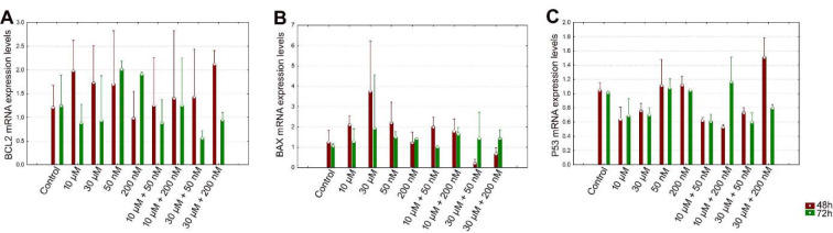
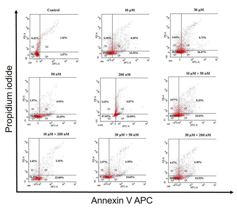
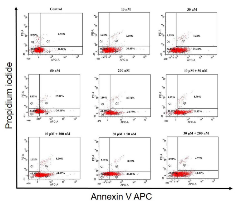
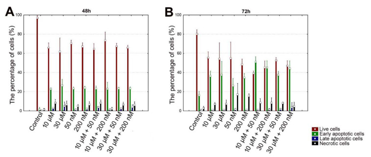
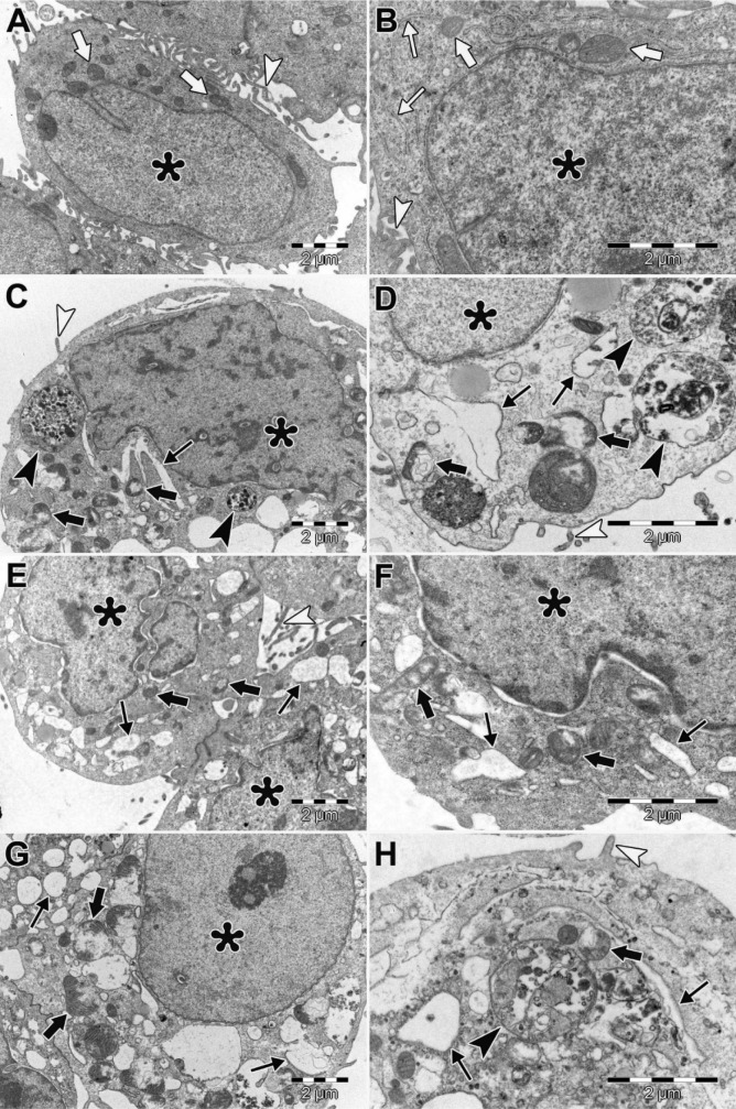
Endometrial cancer is the most common cancer of the female reproductive system. Combination treatment with specific agents has been widely used as a targeted therapy for cancer. In this study, we aimed to investigate the anti-proliferative and apoptotic effects of varying concentrations of perifosine and vitamin D on the human endometrial cancer cell line (HEC-1A). HEC-1A cells were exposed to perifosine (10 μM, 30 μM), vitamin D (50 nM, 200 nM) and combinations of both for 48 h and 72 h. Monitoring of cell proliferation in a time-dependent manner was performed with the xCELLigence RTCA DP system. The levels of BCL2, BAX and P53 mRNA expression were examined using RT-qPCR. Apoptosis was determined using Annexin V, which were followed by flow cytometry analysis. Ultra-structural morphology of cells was analyzed by transmission electron microscopy (TEM) for 72 h. The anti-proliferative and apoptotic effects of the perifosine+vitamin D combination (30 μM + 200 nM at 48 h and 10 μM + 200 nM at 72 h) on HEC-1A cells were higher than in perifosine and vitamin D alone. It was observed that perifosine has increased the expression of BAX mRNA in HEC-1A cells in a dose-dependent manner. While perifosine+vitamin D combinations increased P53 mRNA expression in HEC-1A cells we did not find any significant change in BCL2, BAX mRNA expression levels. In TEM examinations of HEC-1A cells, perifosine appeared to lead autophagic cell death, whereas vitamin D caused paraptosis-like cell death and combination of perifosine+vitamin D caused apoptotic and non-apoptotic (paraptotic, autophagic and necrotic) cell death. Therefore, it is considered that the combination of both drugs in the treatment of endometrial cancer might be an alternative and effective treatment option through activating the apoptotic and non-apoptotic cell death mechanisms in cancer cells.
子宫内膜癌是女性生殖系统最常见的癌症。特定药物的联合治疗已被广泛用作癌症的靶向治疗。在本研究中,我们旨在研究不同浓度的哌立福新和维生素D对人子宫内膜癌细胞系(HEC-1A)的抗增殖和凋亡作用。将HEC-1A细胞暴露于哌立福新(10 μM、30 μM)、维生素D(50 nM、200 nM)以及两者的组合中48小时和72小时。使用xCELLigence RTCA DP系统以时间依赖性方式监测细胞增殖。使用RT-qPCR检测BCL2、BAX和P53 mRNA表达水平。使用膜联蛋白V测定细胞凋亡,随后进行流式细胞术分析。通过透射电子显微镜(TEM)分析细胞72小时的超微结构形态。哌立福新+维生素D组合(48小时时为30 μM + 200 nM,72小时时为10 μM + 200 nM)对HEC-1A细胞的抗增殖和凋亡作用高于单独使用哌立福新和维生素D。观察到哌立福新以剂量依赖性方式增加了HEC-1A细胞中BAX mRNA的表达。虽然哌立福新+维生素D组合增加了HEC-1A细胞中P53 mRNA的表达,但我们未发现BCL2、BAX mRNA表达水平有任何显著变化。在对HEC-1A细胞的TEM检查中,哌立福新似乎导致自噬性细胞死亡,而维生素D导致类副凋亡性细胞死亡,哌立福新+维生素D组合导致凋亡性和非凋亡性(副凋亡性、自噬性和坏死性)细胞死亡。因此,认为两种药物联合用于治疗子宫内膜癌可能是一种通过激活癌细胞凋亡和非凋亡细胞死亡机制的替代且有效的治疗选择。