• 文献检索
  • 文档翻译
  • 深度研究
  • 学术资讯
  • Suppr Zotero 插件Zotero 插件
  • 邀请有礼
  • 套餐&价格
  • 历史记录
应用&插件
Suppr Zotero 插件Zotero 插件浏览器插件Mac 客户端Windows 客户端微信小程序
定价
高级版会员购买积分包购买API积分包
服务
文献检索文档翻译深度研究API 文档MCP 服务
关于我们
关于 Suppr公司介绍联系我们用户协议隐私条款
关注我们

Suppr 超能文献

核心技术专利:CN118964589B侵权必究
粤ICP备2023148730 号-1Suppr @ 2026

文献检索

告别复杂PubMed语法,用中文像聊天一样搜索,搜遍4000万医学文献。AI智能推荐,让科研检索更轻松。

立即免费搜索

文件翻译

保留排版,准确专业,支持PDF/Word/PPT等文件格式,支持 12+语言互译。

免费翻译文档

深度研究

AI帮你快速写综述,25分钟生成高质量综述,智能提取关键信息,辅助科研写作。

立即免费体验

儿童黄斑区形态及视锥细胞密度和间距的活体评估

In vivo assessment of foveal geometry and cone photoreceptor density and spacing in children.

机构信息

College of Optometry, University of Houston, 4901 Calhoun Rd, Houston, TX, 77204-2020, USA.

Department of Biomedical Engineering, University of Houston, 3517 Cullen Blvd, Houston, TX, 77204-5060, USA.

出版信息

Sci Rep. 2020 Jun 2;10(1):8942. doi: 10.1038/s41598-020-65645-2.

DOI:10.1038/s41598-020-65645-2
PMID:32487997
原文链接:https://pmc.ncbi.nlm.nih.gov/articles/PMC7265395/
Abstract

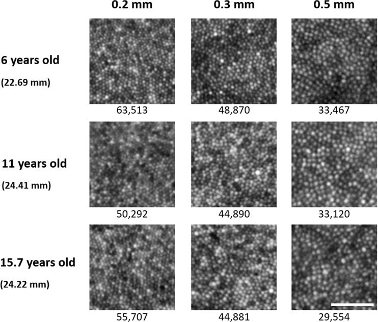
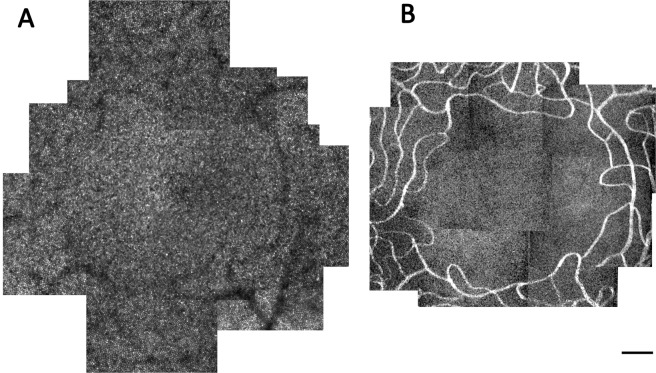
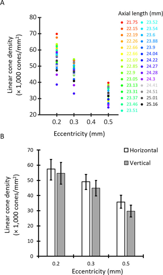
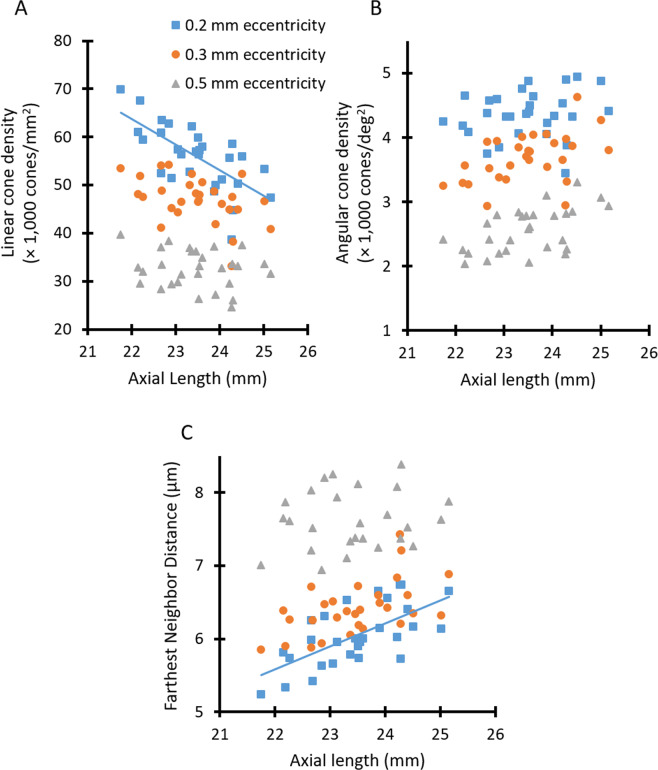
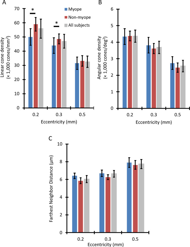
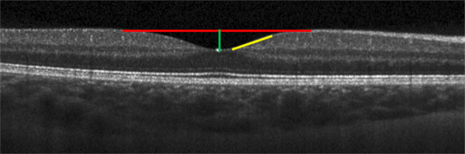
The fovea undergoes significant developmental changes from birth into adolescence. However, there is limited data examining cone photoreceptor density, foveal pit shape, and foveal avascular zone (FAZ) size in children. The purpose of this study was to determine whether overall foveal structure differs as a function of age and refractive status in children. Forty-eight healthy children (ages 5.8 to 15.8 years) underwent optical coherence tomography imaging to quantify foveal point thickness and foveal pit diameter, depth, and slope. Adaptive optics scanning laser ophthalmoscope (AOSLO) images of foveal capillaries and cone photoreceptors were acquired in a subset of children to quantify FAZ metrics and cone densities at 0.2, 0.3, and 0.5 mm eccentricities. Results show that foveal pit and FAZ metrics were not related to age, axial length, or refractive status. However, linear cone density was lower in myopic versus non-myopic children at eccentricities of 0.2 mm (mean ± SD = 50,022 ± 5,878 cones/mm vs 58,989 ± 4,822 cones/mm, P < 0.001) and 0.3 mm (43,944 ± 5,547 cones/mm vs 48,622 ± 3,538 cones/mm, P < 0.001). These results suggest FAZ and foveal pit metrics do not systematically differ with age in children, while myopic eyes have decreased linear cone density near the foveal center. Significance Statement: The development of the fovea begins prior to birth and continues through the early teenage years until it reaches adult-like properties. Although the majority of changes during childhood are related to the maturation and migration of cone photoreceptors, in vivo data describing cone packing in children is limited. We assessed overall foveal structure in children as young as 5.8 years old by quantifying cone density and spacing, foveal avascular zone size, and foveal pit morphometry to investigate potential structural differences as a function of age and refractive status. While foveal avascular zone and foveal pit metrics did not significantly differ with age, results indicate that myopic children have lower linear cone densities close to the foveal center compared to non-myopic children.

摘要

黄斑从出生到青春期经历了显著的发育变化。然而,目前关于儿童的锥体细胞密度、黄斑中心凹形态和黄斑无血管区(FAZ)大小的数据有限。本研究的目的是确定儿童的整体黄斑结构是否随年龄和屈光状态的不同而不同。48 名健康儿童(年龄 5.8 至 15.8 岁)接受光学相干断层扫描成像,以量化黄斑中心凹点厚度和黄斑中心凹直径、深度和斜率。在一部分儿童中,使用自适应光学扫描激光检眼镜(AOSLO)获取黄斑毛细血管和锥体细胞图像,以量化 FAZ 指标和 0.2、0.3 和 0.5mm 偏心处的锥体细胞密度。结果表明,黄斑中心凹形态和 FAZ 指标与年龄、眼轴长度或屈光状态无关。然而,在 0.2mm(平均±SD=50022±5878 个/毫米与 58989±4822 个/毫米,P<0.001)和 0.3mm(43944±5547 个/毫米与 48622±3538 个/毫米,P<0.001)偏心处,近视儿童的线性锥体细胞密度低于非近视儿童。这些结果表明,FAZ 和黄斑中心凹形态在儿童中不会随年龄系统地变化,而近视眼在黄斑中心附近的线性锥体细胞密度降低。

意义声明

黄斑的发育始于出生前,并持续到青少年早期,直到达到成人样特征。虽然儿童时期的大多数变化与锥体细胞的成熟和迁移有关,但描述儿童锥体细胞排列的数据有限。我们通过量化锥体细胞密度和间距、FAZ 大小以及黄斑中心凹形态测量值,评估了年龄在 5.8 岁以下的儿童的整体黄斑结构,以研究年龄和屈光状态对其潜在结构的影响。虽然 FAZ 和黄斑中心凹形态指标与年龄无显著差异,但结果表明,与非近视儿童相比,近视儿童在黄斑中心附近的线性锥体细胞密度较低。

https://cdn.ncbi.nlm.nih.gov/pmc/blobs/e0f9/7265395/eaa408a21eee/41598_2020_65645_Fig6_HTML.jpg
https://cdn.ncbi.nlm.nih.gov/pmc/blobs/e0f9/7265395/688679e79d51/41598_2020_65645_Fig1_HTML.jpg
https://cdn.ncbi.nlm.nih.gov/pmc/blobs/e0f9/7265395/1ebdbeb86e79/41598_2020_65645_Fig2_HTML.jpg
https://cdn.ncbi.nlm.nih.gov/pmc/blobs/e0f9/7265395/de72da4a7899/41598_2020_65645_Fig3_HTML.jpg
https://cdn.ncbi.nlm.nih.gov/pmc/blobs/e0f9/7265395/b52f25be4a07/41598_2020_65645_Fig4_HTML.jpg
https://cdn.ncbi.nlm.nih.gov/pmc/blobs/e0f9/7265395/55d235fb11a0/41598_2020_65645_Fig5_HTML.jpg
https://cdn.ncbi.nlm.nih.gov/pmc/blobs/e0f9/7265395/eaa408a21eee/41598_2020_65645_Fig6_HTML.jpg

相似文献

1
In vivo assessment of foveal geometry and cone photoreceptor density and spacing in children.儿童黄斑区形态及视锥细胞密度和间距的活体评估
Sci Rep. 2020 Jun 2;10(1):8942. doi: 10.1038/s41598-020-65645-2.
2
The association between the foveal avascular zone and retinal thickness.黄斑无血管区与视网膜厚度之间的关联。
Invest Ophthalmol Vis Sci. 2014 Sep 30;55(10):6870-7. doi: 10.1167/iovs.14-15446.
3
Imaging Foveal Microvasculature: Optical Coherence Tomography Angiography Versus Adaptive Optics Scanning Light Ophthalmoscope Fluorescein Angiography.黄斑区微血管成像:光学相干断层扫描血管造影术与自适应光学扫描激光眼底镜荧光血管造影术的对比
Invest Ophthalmol Vis Sci. 2016 Jul 1;57(9):OCT130-40. doi: 10.1167/iovs.15-18932.
4
Relationship between the foveal avascular zone and foveal pit morphology.中心凹无血管区与中心凹小凹形态的关系。
Invest Ophthalmol Vis Sci. 2012 Mar 21;53(3):1628-36. doi: 10.1167/iovs.11-8488. Print 2012 Mar.
5
Foveal avascular zone and its relationship to foveal pit shape.黄斑无血管区及其与黄斑中心凹形态的关系。
Optom Vis Sci. 2012 May;89(5):602-10. doi: 10.1097/OPX.0b013e3182504227.
6
The Influence of Myopia on the Foveal Avascular Zone and Density of Blood Vessels of the Macula-An OCTA Study.近视对黄斑中心凹无血管区及脉络膜血管密度的影响——OCTA 研究。
Medicina (Kaunas). 2023 Feb 24;59(3):452. doi: 10.3390/medicina59030452.
7
Intersubject variability of foveal cone photoreceptor density in relation to eye length.与眼轴长度相关的黄斑中心凹锥体细胞密度的个体间变异性。
Invest Ophthalmol Vis Sci. 2010 Dec;51(12):6858-67. doi: 10.1167/iovs.10-5499. Epub 2010 Aug 4.
8
Normative Data Assessment of Peripapillary and Macular Vessel Density and Foveal Avascular Zone Metrics Using Optical Coherence Tomography Angiography in Children.使用光学相干断层血管造影术评估儿童视乳头周围和黄斑血管密度及中心无血管区参数的正常值。
J Pediatr Ophthalmol Strabismus. 2020 Nov 1;57(6):388-398. doi: 10.3928/01913913-20200903-01.
9
Human foveal cone photoreceptor topography and its dependence on eye length.人眼中心凹锥光感受器的分布及其与眼轴长度的关系。
Elife. 2019 Jul 26;8:e47148. doi: 10.7554/eLife.47148.
10
Optical coherence tomography angiography of superficial retinal vessel density and foveal avascular zone in myopic children.近视儿童的浅层视网膜血管密度和中心无血管区的光相干断层扫描血管造影。
PLoS One. 2019 Jul 18;14(7):e0219785. doi: 10.1371/journal.pone.0219785. eCollection 2019.

引用本文的文献

1
In vivo assessment of cone loss and macular perfusion in children with myopia.近视儿童的锥体损失和黄斑灌注的体内评估。
Sci Rep. 2024 Nov 2;14(1):26373. doi: 10.1038/s41598-024-78280-y.
2
The effect of sampling window size on topographical maps of foveal cone density.采样窗口大小对中央凹视锥细胞密度地形图的影响。
Front Ophthalmol (Lausanne). 2024 Apr 9;4:1348950. doi: 10.3389/fopht.2024.1348950. eCollection 2024.
3
Adaptive Optics Retinal Imaging in RDH12-Associated Early Onset Severe Retinal Dystrophy.RDH12相关早发性严重视网膜营养不良的自适应光学视网膜成像

本文引用的文献

1
Assessment of Macular Microvasculature in Healthy Eyes of Infants and Children Using OCT Angiography.利用 OCT 血管造影评估婴儿和儿童健康眼中的黄斑微循环。
Ophthalmology. 2019 Dec;126(12):1703-1711. doi: 10.1016/j.ophtha.2019.06.028. Epub 2019 Jul 15.
2
Optical coherence tomography angiography of superficial retinal vessel density and foveal avascular zone in myopic children.近视儿童的浅层视网膜血管密度和中心无血管区的光相干断层扫描血管造影。
PLoS One. 2019 Jul 18;14(7):e0219785. doi: 10.1371/journal.pone.0219785. eCollection 2019.
3
High-resolution imaging of photoreceptors in healthy human eyes using an adaptive optics retinal camera.
Invest Ophthalmol Vis Sci. 2024 Mar 5;65(3):9. doi: 10.1167/iovs.65.3.9.
4
Cone Density Is Correlated to Outer Segment Length and Retinal Thickness in the Human Foveola.人中央凹的 cones 密度与 outer segment length 和视网膜厚度相关。
Invest Ophthalmol Vis Sci. 2023 Dec 1;64(15):11. doi: 10.1167/iovs.64.15.11.
5
IMI-Nonpathological Human Ocular Tissue Changes With Axial Myopia.轴性近视的非病理性人眼球组织变化。
Invest Ophthalmol Vis Sci. 2023 May 1;64(6):5. doi: 10.1167/iovs.64.6.5.
6
A variation of foveal morphology in a group of children with hypermetropia.一组远视儿童的黄斑形态变异。
Int Ophthalmol. 2023 Aug;43(8):2947-2956. doi: 10.1007/s10792-023-02701-6. Epub 2023 Apr 20.
7
Macular outer nuclear layer, ellipsoid zone and outer photoreceptor segment band thickness, axial length and other determinants.黄斑外核层、椭圆体带和外光感受器节段带厚度、眼轴长度和其他决定因素。
Sci Rep. 2023 Apr 3;13(1):5386. doi: 10.1038/s41598-023-32629-x.
8
Twenty-five years of clinical applications using adaptive optics ophthalmoscopy [Invited].使用自适应光学检眼镜的25年临床应用[特邀报告]
Biomed Opt Express. 2022 Dec 20;14(1):387-428. doi: 10.1364/BOE.472274. eCollection 2023 Jan 1.
9
Photoreceptor density in relation to axial length and retinal location in human eyes.人眼的光感受器密度与眼轴长度和视网膜位置的关系。
Sci Rep. 2022 Dec 9;12(1):21371. doi: 10.1038/s41598-022-25460-3.
10
Retinal magnification factors at the fixation locus derived from schematic eyes with four individualized surfaces.源自具有四个个性化表面的简化眼在注视位点处的视网膜放大率因子。
Biomed Opt Express. 2022 Jun 8;13(7):3786-3808. doi: 10.1364/BOE.460553. eCollection 2022 Jul 1.
利用自适应光学视网膜相机对健康人眼的光感受器进行高分辨率成像。
Eye (Lond). 2018 Nov;32(11):1723-1730. doi: 10.1038/s41433-018-0140-1. Epub 2018 Jul 11.
4
Quantitative retinal microvasculature in children using swept-source optical coherence tomography: the Hong Kong Children Eye Study.利用扫频源光学相干断层扫描技术对儿童视网膜微血管进行定量分析:香港儿童眼病研究
Br J Ophthalmol. 2018 Jun 28. doi: 10.1136/bjophthalmol-2018-312413.
5
Factors affecting foveal avascular zone in healthy eyes: An examination using swept-source optical coherence tomography angiography.健康眼睛中影响黄斑无血管区的因素:使用扫频源光学相干断层扫描血管造影术的检查
PLoS One. 2017 Nov 27;12(11):e0188572. doi: 10.1371/journal.pone.0188572. eCollection 2017.
6
Distribution differences of macular cones measured by AOSLO: Variation in slope from fovea to periphery more pronounced than differences in total cones.通过自适应光学扫描激光眼底镜测量的黄斑视锥细胞分布差异:从中央凹到周边的斜率变化比视锥细胞总数的差异更明显。
Vision Res. 2017 Mar;132:62-68. doi: 10.1016/j.visres.2016.06.015. Epub 2016 Nov 3.
7
Evaluating Descriptive Metrics of the Human Cone Mosaic.评估人类视锥细胞镶嵌的描述性指标。
Invest Ophthalmol Vis Sci. 2016 Jun 1;57(7):2992-3001. doi: 10.1167/iovs.16-19072.
8
Global Prevalence of Myopia and High Myopia and Temporal Trends from 2000 through 2050.全球近视和高度近视的患病率及 2000 至 2050 年的时间趋势。
Ophthalmology. 2016 May;123(5):1036-42. doi: 10.1016/j.ophtha.2016.01.006. Epub 2016 Feb 11.
9
In Vivo Foveal Development Using Optical Coherence Tomography.利用光学相干断层扫描技术进行的活体黄斑中心凹发育研究
Invest Ophthalmol Vis Sci. 2015 Jul;56(8):4537-45. doi: 10.1167/iovs.15-16542.
10
Macular retinal layer thickness in childhood.儿童期黄斑视网膜层厚度
Retina. 2015 Jun;35(6):1223-33. doi: 10.1097/IAE.0000000000000464.