• 文献检索
  • 文档翻译
  • 深度研究
  • 学术资讯
  • Suppr Zotero 插件Zotero 插件
  • 邀请有礼
  • 套餐&价格
  • 历史记录
应用&插件
Suppr Zotero 插件Zotero 插件浏览器插件Mac 客户端Windows 客户端微信小程序
定价
高级版会员购买积分包购买API积分包
服务
文献检索文档翻译深度研究API 文档MCP 服务
关于我们
关于 Suppr公司介绍联系我们用户协议隐私条款
关注我们

Suppr 超能文献

核心技术专利:CN118964589B侵权必究
粤ICP备2023148730 号-1Suppr @ 2026

文献检索

告别复杂PubMed语法,用中文像聊天一样搜索,搜遍4000万医学文献。AI智能推荐,让科研检索更轻松。

立即免费搜索

文件翻译

保留排版,准确专业,支持PDF/Word/PPT等文件格式,支持 12+语言互译。

免费翻译文档

深度研究

AI帮你快速写综述,25分钟生成高质量综述,智能提取关键信息,辅助科研写作。

立即免费体验

DryMass:处理和分析球形、细胞大小物体的定量相显微镜图像。

DryMass: handling and analyzing quantitative phase microscopy images of spherical, cell-sized objects.

机构信息

Biotechnology Center, Center for Molecular and Cellular Bioengineering, Technische Universität Dresden, Tatzberg 47/49, Dresden, 01307, Germany.

Max Planck Institute for the Science of Light, Max-Planck-Zentrum für Physik und Medizin, Staudtstr. 2, Erlangen, 91058, Germany.

出版信息

BMC Bioinformatics. 2020 Jun 3;21(1):226. doi: 10.1186/s12859-020-03553-y.

DOI:10.1186/s12859-020-03553-y
PMID:32493205
原文链接:https://pmc.ncbi.nlm.nih.gov/articles/PMC7268593/
Abstract

BACKGROUND

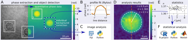
Quantitative phase imaging (QPI) is an established tool for the marker-free classification and quantitative characterization of biological samples. For spherical objects, such as cells in suspension, microgel beads, or liquid droplets, a single QPI image is sufficient to extract the radius and the average refractive index. This technique is invaluable, as it allows the characterization of large sample populations at high measurement rates. However, until now, no universal software existed that could perform this type of analysis. Besides the choice of imaging modality and the variety in imaging software, the main difficulty has been to automate the entire analysis pipeline from raw data to ensemble statistics.

RESULTS

We present DryMass, a powerful tool for QPI that covers all relevant steps from loading experimental data (multiple file formats supported), computing the phase data (built-in, automated hologram analysis), performing phase background corrections (offset, tilt, second order polynomial) to fitting scattering models (light projection, Rytov approximation, Mie simulations) to spherical phase objects for the extraction of dry mass, radius, and average refractive index. The major contribution of DryMass is a user-convenient, reliable, reproducible, and automated analysis pipeline for an arbitrary number of QPI datasets of arbitrary sizes.

CONCLUSION

DryMass is a leap forward for data analysis in QPI, as it not only makes it easier to visualize raw QPI data and reproduce previous results in the field, but it also opens up QPI analysis to users without a background in programming or phase imaging.

摘要

背景

定量相位成像(QPI)是一种用于对生物样本进行无标记分类和定量特征描述的成熟工具。对于球形物体,如悬浮细胞、微凝胶珠或液滴,单个 QPI 图像足以提取半径和平均折射率。这项技术非常有价值,因为它可以在高测量速率下对大量样本进行特征描述。然而,到目前为止,还没有通用的软件可以执行这种类型的分析。除了成像方式的选择和成像软件的多样性之外,主要的困难是将从原始数据到总体统计的整个分析流程自动化。

结果

我们提出了 DryMass,这是一款用于 QPI 的强大工具,涵盖了从加载实验数据(支持多种文件格式)、计算相位数据(内置、自动全息分析)、执行相位背景校正(偏移、倾斜、二次多项式)到拟合散射模型(光投影、Rytov 近似、Mie 模拟)到球形相位物体的所有相关步骤,以提取干质量、半径和平均折射率。DryMass 的主要贡献是为任意数量的任意大小的 QPI 数据集提供了一个用户友好、可靠、可重复且自动化的分析流程。

结论

DryMass 是 QPI 数据分析的一个飞跃,因为它不仅使可视化原始 QPI 数据和重现该领域以前的结果变得更加容易,而且还为没有编程或相位成像背景的用户打开了 QPI 分析的大门。

https://cdn.ncbi.nlm.nih.gov/pmc/blobs/9ff9/7268593/48553315ae4c/12859_2020_3553_Fig3_HTML.jpg
https://cdn.ncbi.nlm.nih.gov/pmc/blobs/9ff9/7268593/f20c3c8dce7c/12859_2020_3553_Fig1_HTML.jpg
https://cdn.ncbi.nlm.nih.gov/pmc/blobs/9ff9/7268593/7665b41bd21d/12859_2020_3553_Fig2_HTML.jpg
https://cdn.ncbi.nlm.nih.gov/pmc/blobs/9ff9/7268593/48553315ae4c/12859_2020_3553_Fig3_HTML.jpg
https://cdn.ncbi.nlm.nih.gov/pmc/blobs/9ff9/7268593/f20c3c8dce7c/12859_2020_3553_Fig1_HTML.jpg
https://cdn.ncbi.nlm.nih.gov/pmc/blobs/9ff9/7268593/7665b41bd21d/12859_2020_3553_Fig2_HTML.jpg
https://cdn.ncbi.nlm.nih.gov/pmc/blobs/9ff9/7268593/48553315ae4c/12859_2020_3553_Fig3_HTML.jpg

相似文献

1
DryMass: handling and analyzing quantitative phase microscopy images of spherical, cell-sized objects.DryMass:处理和分析球形、细胞大小物体的定量相显微镜图像。
BMC Bioinformatics. 2020 Jun 3;21(1):226. doi: 10.1186/s12859-020-03553-y.
2
Accurate evaluation of size and refractive index for spherical objects in quantitative phase imaging.定量相成像中球形物体尺寸和折射率的精确评估。
Opt Express. 2018 Apr 16;26(8):10729-10743. doi: 10.1364/OE.26.010729.
3
Refractive index measurements of single, spherical cells using digital holographic microscopy.使用数字全息显微镜对单个球形细胞进行折射率测量。
Methods Cell Biol. 2015;125:143-59. doi: 10.1016/bs.mcb.2014.10.016. Epub 2015 Jan 8.
4
CellSNAP: a fast, accurate algorithm for 3D cell segmentation in quantitative phase imaging.CellSNAP:一种用于定量相衬成像中三维细胞分割的快速、准确算法。
J Biomed Opt. 2024 Jun;29(Suppl 2):S22706. doi: 10.1117/1.JBO.29.S2.S22706. Epub 2024 Apr 18.
5
Quantitative phase imaging for cell culture quality control.用于细胞培养质量控制的定量相成像
Cytometry A. 2017 May;91(5):470-481. doi: 10.1002/cyto.a.23082. Epub 2017 Mar 6.
6
Accurate single-shot quantitative phase imaging of biological specimens with telecentric digital holographic microscopy.远心数字全息显微镜对生物样本进行精确的单次定量相位成像。
J Biomed Opt. 2014 Apr;19(4):046022. doi: 10.1117/1.JBO.19.4.046022.
7
Robust Phase Unwrapping via Deep Image Prior for Quantitative Phase Imaging.基于深度图像先验的定量相位成像稳健相位解包裹。
IEEE Trans Image Process. 2021;30:7025-7037. doi: 10.1109/TIP.2021.3099956. Epub 2021 Aug 10.
8
Quantitative phase imaging techniques for measuring scattering properties of cells and tissues: a review-part I.定量相位成像技术测量细胞和组织的散射特性:综述第一部分。
J Biomed Opt. 2024 Jun;29(Suppl 2):S22713. doi: 10.1117/1.JBO.29.S2.S22713. Epub 2024 Jul 18.
9
Quantitative Phase Imaging: Recent Advances and Expanding Potential in Biomedicine.定量相位成像:生物医学中的最新进展和扩展潜力。
ACS Nano. 2022 Aug 23;16(8):11516-11544. doi: 10.1021/acsnano.1c11507. Epub 2022 Aug 2.
10
Integral refractive index determination of living suspension cells by multifocus digital holographic phase contrast microscopy.利用多焦点数字全息相衬显微镜测定活悬浮细胞的积分折射率
J Biomed Opt. 2007 Sep-Oct;12(5):054009. doi: 10.1117/1.2798639.

引用本文的文献

1
Fabrication and validation of an LED array microscope for multimodal, quantitative imaging.用于多模态定量成像的LED阵列显微镜的制造与验证
HardwareX. 2023 Jan 24;13:e00399. doi: 10.1016/j.ohx.2023.e00399. eCollection 2023 Mar.
2
HoloPhaseNet: fully automated deep-learning-based hologram reconstruction using a conditional generative adversarial model.全相位网络:基于条件生成对抗模型的全自动深度学习全息图重建
Biomed Opt Express. 2022 Jun 27;13(7):4032-4046. doi: 10.1364/BOE.452645. eCollection 2022 Jul 1.
3
PNIPAAm microgels with defined network architecture as temperature sensors in optical stretchers.

本文引用的文献

1
Standardized microgel beads as elastic cell mechanical probes.标准化微凝胶珠作为弹性细胞力学探针。
J Mater Chem B. 2018 Oct 21;6(39):6245-6261. doi: 10.1039/c8tb01421c. Epub 2018 Sep 13.
2
Accurate evaluation of size and refractive index for spherical objects in quantitative phase imaging.定量相成像中球形物体尺寸和折射率的精确评估。
Opt Express. 2018 Apr 16;26(8):10729-10743. doi: 10.1364/OE.26.010729.
3
Three-dimensional correlative single-cell imaging utilizing fluorescence and refractive index tomography.利用荧光和折射率断层扫描的三维相关单细胞成像。
具有确定网络结构的聚N-异丙基丙烯酰胺微凝胶作为光镊中的温度传感器。
Mater Adv. 2022 Jul 5;3(15):6179-6190. doi: 10.1039/d2ma00296e. eCollection 2022 Aug 1.
4
Multiparametric quantitative phase imaging for real-time, single cell, drug screening in breast cancer.用于实时、单细胞、乳腺癌药物筛选的多参数定量相位成像。
Commun Biol. 2022 Aug 8;5(1):794. doi: 10.1038/s42003-022-03759-1.
5
Quantitative Phase Imaging: Recent Advances and Expanding Potential in Biomedicine.定量相位成像:生物医学中的最新进展和扩展潜力。
ACS Nano. 2022 Aug 23;16(8):11516-11544. doi: 10.1021/acsnano.1c11507. Epub 2022 Aug 2.
6
Spatio-temporal performance in an incoherent holography lattice light-sheet microscope (IHLLS).非相干全息晶格光片显微镜(IHLLS)中的时空性能。
Opt Express. 2021 Jul 19;29(15):23888-23901. doi: 10.1364/OE.425069.
J Biophotonics. 2018 Mar;11(3). doi: 10.1002/jbio.201700145. Epub 2017 Sep 25.
4
Cell nuclei have lower refractive index and mass density than cytoplasm.细胞核的折射率和质量密度低于细胞质。
J Biophotonics. 2016 Oct;9(10):1068-1076. doi: 10.1002/jbio.201500273. Epub 2016 Mar 24.
5
Diffraction optical tomography using a quantitative phase imaging unit.使用定量相位成像单元的衍射光学层析成像。
Opt Lett. 2014 Dec 15;39(24):6935-8. doi: 10.1364/OL.39.006935.
6
scikit-image: image processing in Python.scikit-image:在 Python 中进行图像处理。
PeerJ. 2014 Jun 19;2:e453. doi: 10.7717/peerj.453. eCollection 2014.
7
Digital holographic microtomography for high-resolution refractive index mapping of live cells.数字全息显微层析术用于活细胞的高分辨率折射率映射。
J Biophotonics. 2013 May;6(5):416-24. doi: 10.1002/jbio.201200022. Epub 2012 Aug 24.
8
Fiji: an open-source platform for biological-image analysis.斐济:一个用于生物影像分析的开源平台。
Nat Methods. 2012 Jun 28;9(7):676-82. doi: 10.1038/nmeth.2019.
9
Noninvasive time-dependent cytometry monitoring by digital holography.通过数字全息术进行无创的时间依赖性细胞计数监测
J Biomed Opt. 2007 Nov-Dec;12(6):064002. doi: 10.1117/1.2804926.
10
Integral refractive index determination of living suspension cells by multifocus digital holographic phase contrast microscopy.利用多焦点数字全息相衬显微镜测定活悬浮细胞的积分折射率
J Biomed Opt. 2007 Sep-Oct;12(5):054009. doi: 10.1117/1.2798639.