Cañizares Gabriel, Gonzalez-Montoro Andrea, Freire Marta, Lamprou Efthymios, Barrio John, Sanchez Filomeno, Benlloch José M, Hernandez Liczandro, Moliner Laura, Vidal Luis F, Torres Irene, Sopena Pablo, Vera-Donoso Cesar D, Bello Pilar, Barbera Julio, Gonzalez Antonio J
Instituto de Instrumentación para Imagen Molecular (I3M), Centro Mixto CSIC - Universitat Politècnica de València, Camino de Vera s/n, 46022, Valencia, Spain.
Servicio de Medicina Nuclear, Área Clínica de Imagen Médica, Hospital Univ. y Polit. La Fe, 46026, Valencia, Spain.
EJNMMI Phys. 2020 Jun 5;7(1):38. doi: 10.1186/s40658-020-00305-y.
Prostate cancer (PCa) represents one of the most common types of cancers facing the male population. Nowadays, to confirm PCa, systematic or multiparametric MRI-targeted transrectal or transperineal biopsies of the prostate are required. However, due to the lack of an accurate imaging technique capable to precisely locate cancerous cells in the prostate, ultrasound biopsies sample random parts of the prostate and, therefore, it is possible to miss regions where those cancerous cells are present. In spite of the improvement with multiparametric MRI, the low reproducibility of its reading undermines the specificity of the method. Recent development of prostate-specific radiotracers has grown the interest on using positron emission tomography (PET) scanners for this purpose, but technological improvements are still required (current scanners have resolutions in the range of 4-5 mm).
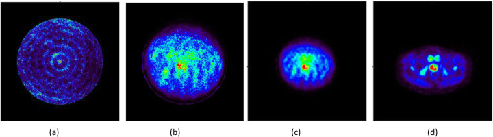
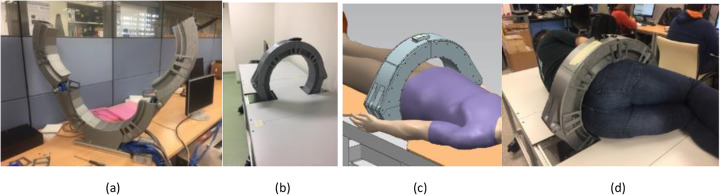
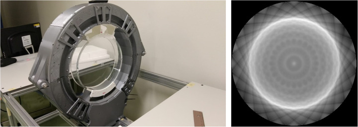
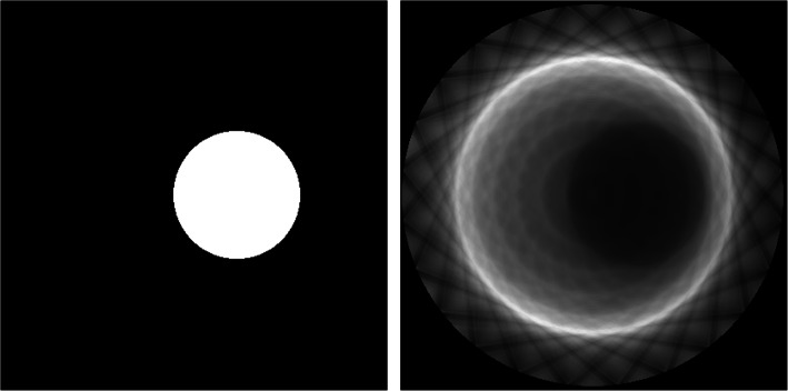
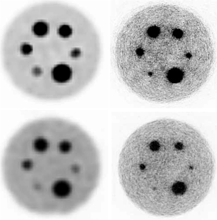
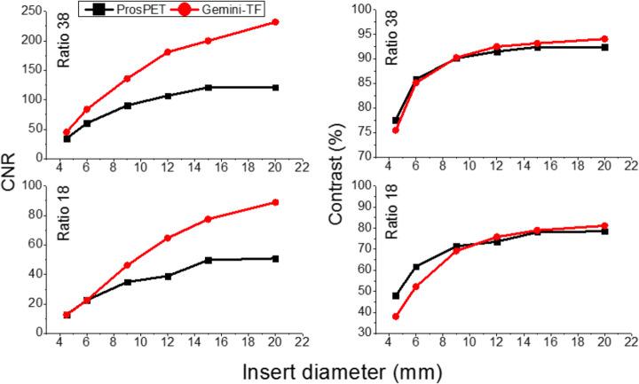
The main goal of this work is to improve state-of-the-art PCa imaging and diagnosis. We have focused our efforts on the design of a novel prostate-dedicated PET scanner, named ProsPET. This system has small scanner dimensions defined by a ring of just 41 cm inner diameter. In this work, we report the design, implementation, and evaluation (both through simulations and real data) of the ProsPET scanner. We have been able to achieve < 2 mm resolution in reconstructed images and high sensitivity. In addition, we have included a comparison with the Philips Gemini-TF scanner, which is used for routine imaging of PCa patients. The ProsPET exhibits better contrast, especially for rod sizes as small as 4.5 mm in diameter. Finally, we also show the first reconstructed image of a PCa patient acquired with the ProsPET.
We have designed and built a prostate specific PET system, with a small footprint and improved spatial resolution when compared to conventional whole-body PET scanners. The gamma ray impact within each detector block includes accurate DOI determination, correcting for the parallax error. The potential role of combined organ-dedicated prostate-specific membrane antigen (PSMA) PET and ultrasound devices, as a prebiopsy diagnostic tool, could be used to guide sampling of the most aggressive sites in the prostate.
前列腺癌(PCa)是男性群体面临的最常见癌症类型之一。如今,要确诊前列腺癌,需要对前列腺进行系统性或多参数MRI靶向经直肠或经会阴活检。然而,由于缺乏能够精确在前列腺中定位癌细胞的准确成像技术,超声活检对前列腺的随机部位进行采样,因此有可能遗漏存在癌细胞的区域。尽管多参数MRI有所改进,但其读数的低重复性削弱了该方法的特异性。前列腺特异性放射性示踪剂的最新发展增加了为此目的使用正电子发射断层扫描(PET)扫描仪的兴趣,但仍需要技术改进(当前扫描仪的分辨率在4 - 5毫米范围内)。
这项工作的主要目标是改进当前最先进的前列腺癌成像和诊断。我们将精力集中在设计一种名为ProsPET的新型前列腺专用PET扫描仪上。该系统具有小尺寸的扫描仪,内径仅为41厘米的环定义了其大小。在这项工作中,我们报告了ProsPET扫描仪的设计、实施以及评估(通过模拟和真实数据)。我们在重建图像中能够实现小于2毫米的分辨率以及高灵敏度。此外,我们还与用于前列腺癌患者常规成像的飞利浦Gemini - TF扫描仪进行了比较。ProsPET表现出更好的对比度,特别是对于直径小至4.5毫米的棒状物体。最后,我们还展示了使用ProsPET获取的前列腺癌患者的首张重建图像。
我们设计并构建了一种前列腺特异性PET系统,与传统的全身PET扫描仪相比,其占地面积小且空间分辨率有所提高。每个探测器模块内的伽马射线影响包括准确的深度-of-interaction(DOI)测定,用于校正视差误差。联合器官专用前列腺特异性膜抗原(PSMA)PET和超声设备作为活检前诊断工具的潜在作用,可用于指导对前列腺中最具侵袭性部位的采样。