Clayson Keyton, Pavlatos Elias, Pan Xueliang, Sandwisch Thomas, Ma Yanhui, Liu Jun
Department of Biomedical Engineering, The Ohio State University, Columbus, OH, USA.
Biophysics Interdisciplinary Group, The Ohio State University, Columbus, OH, USA.
Transl Vis Sci Technol. 2020 Jan 30;9(1):5. doi: 10.1167/tvst.9.1.5. eCollection 2020 Jan.
In vivo evaluation of corneal biomechanics holds the potential for improving diagnosis and management of ocular diseases. We aimed to develop an ocular pulse elastography (OPE) technique to quantify corneal strains generated by naturally occurring pulsations of the intraocular pressure (IOP) using high-frequency ultrasound.
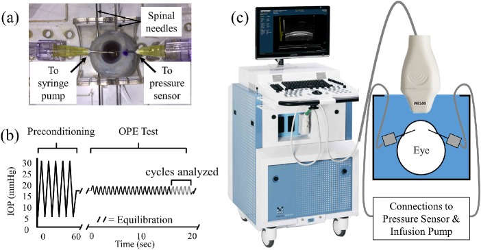
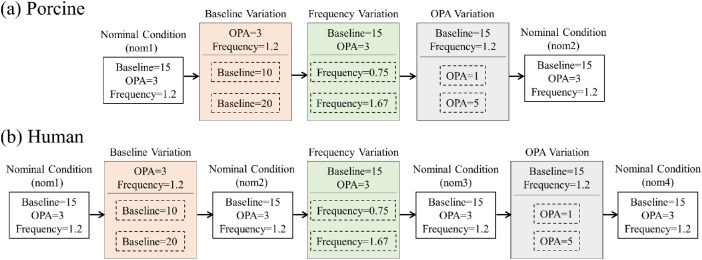
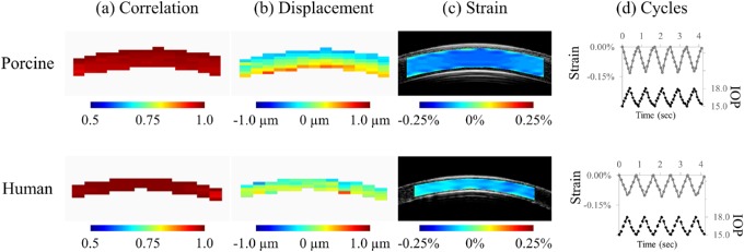
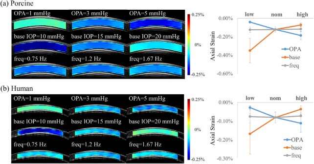
Simulated ocular pulses were induced in whole porcine and human donor globes to investigate the effects of physiologic variations in baseline IOP, ocular pulse amplitude, and frequency on corneal strains. Ocular pulse-induced strains were measured in additional globes before and after UVA-riboflavin-induced corneal crosslinking. The central cornea in each eye was imaged with a 50-MHz ultrasound imaging system and correlation-based speckle tracking of radiofrequency data was used to calculate tissue displacements and strains.
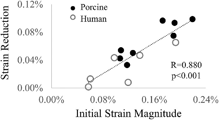
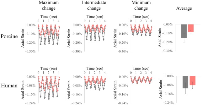
Ocular pulse-induced corneal strains followed the cyclic changes of IOP. Both baseline IOP and ocular pulse amplitude had a significant influence on strain magnitude. Variations in pulse frequency within the normal human heart rate range did not introduce detectable changes in corneal strains. A significant decrease of corneal strain, as quantified by the OPE technique, was observed after corneal crosslinking. The extent of corneal stiffening (i.e., strain reduction) seemed to correlate with the initial strain magnitude.
This ex vivo study demonstrated the feasibility of the OPE method to quantify corneal strains generated by IOP pulsation and detect changes associated with corneal crosslinking treatment.
Integrating in vivo measurement of IOP and ocular pulse amplitude, the OPE method may lead to a new clinical tool for safe and quick biomechanical evaluations of the cornea.
角膜生物力学的体内评估具有改善眼部疾病诊断和管理的潜力。我们旨在开发一种眼部脉搏弹性成像(OPE)技术,以使用高频超声量化由眼内压(IOP)的自然脉动产生的角膜应变。
在整个猪眼和人类供体眼球中诱导模拟眼部脉搏,以研究基线IOP、眼部脉搏幅度和频率的生理变化对角膜应变的影响。在紫外线-核黄素诱导的角膜交联前后,在另外的眼球中测量眼部脉搏诱导的应变。用50-MHz超声成像系统对每只眼睛的中央角膜进行成像,并使用基于相关性的射频数据斑点追踪来计算组织位移和应变。
眼部脉搏诱导的角膜应变跟随IOP的周期性变化。基线IOP和眼部脉搏幅度均对应变幅度有显著影响。在正常人类心率范围内的脉搏频率变化未引起角膜应变的可检测变化。角膜交联后,通过OPE技术量化观察到角膜应变显著降低。角膜变硬的程度(即应变降低)似乎与初始应变幅度相关。
这项离体研究证明了OPE方法量化由IOP脉动产生的角膜应变并检测与角膜交联治疗相关变化的可行性。
整合IOP和眼部脉搏幅度的体内测量,OPE方法可能会导致一种用于角膜安全快速生物力学评估的新临床工具。