Nath Audrey, Robinson Meghan, Magnotti John, Karas Patrick, Curry Daniel, Paldino Michael
Department of Pediatric Neurology, Baylor College of Medicine, Houston, TX, USA.
Core for Advanced MRI, Baylor College of Medicine, Houston, TX, USA.
J Epilepsy Res. 2019 Dec 31;9(2):93-102. doi: 10.14581/jer.19011. eCollection 2019 Dec.
The current tools available for localization of expressive language, including functional magnetic resonance imaging (fMRI) and cortical stimulation mapping (CSM), require that the patient remain stationary and follow language commands with precise timing. Many pediatric epilepsy patients, however, have intact language skills but are unable to participate in these tasks due to cognitive impairments or young age. In adult subjects, there is evidence that language laterality can be determined by resting state (RS) fMRI activity, however there are few studies on the use of RS to accurately predict language laterality in children.
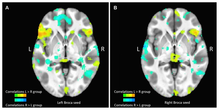
A retrospective review of pediatric patients at Texas Children's Hospital was performed to identify patients who have undergone epilepsy surgical planning over 3 years with language localization using traditional methods of Wada testing, CSM, or task-based fMRI with calculated laterality index, as well as a 7-minute RS scan available without excessive motion or noise. We found the correlation between each subject's left and right Broca's region activity and each of 68 cortical regions.
A group of nine patients with left-lateralized language were found to have greater voxel-wise correlations than a group of six patients with right-lateralized language between a left hemispheric Broca's region seed and the following six cortical regions: left inferior temporal, left lateral orbitofrontal, left pars triangularis, right lateral orbitofrontal, right pars orbitalis and right superior frontal regions.
In a cohort of children with epilepsy, we found that patients with left- and right-hemispheric language lateralization have different RS networks.
目前用于定位表达性语言的工具,包括功能磁共振成像(fMRI)和皮质刺激图谱(CSM),要求患者保持静止并精确计时地遵循语言指令。然而,许多小儿癫痫患者虽具备完整的语言技能,但由于认知障碍或年龄较小而无法参与这些任务。在成年受试者中,有证据表明语言优势半球可通过静息态(RS)fMRI活动来确定,然而关于使用RS准确预测儿童语言优势半球的研究却很少。
对德克萨斯儿童医院的小儿患者进行回顾性研究,以确定那些在3年多时间里接受过癫痫手术规划的患者,这些患者使用传统的Wada测试、CSM或基于任务的fMRI进行语言定位,并计算了优势半球指数,同时还进行了一次7分钟的RS扫描,扫描过程中无过度运动或噪音。我们发现了每个受试者左右布洛卡区活动与68个皮质区域中每个区域之间的相关性。
发现一组9名语言优势半球在左侧的患者,与一组6名语言优势半球在右侧的患者相比,左侧半球布洛卡区种子点与以下六个皮质区域之间的体素相关性更强:左侧颞下回、左侧眶额外侧、左侧三角部、右侧眶额外侧、右侧眶部和右侧额上回。
在一组癫痫患儿中,我们发现语言优势半球在左侧和右侧的患者具有不同的RS网络。