Department of Pathology, Johns Hopkins University School of Medicine, Baltimore, Maryland.
Department of Pathology, Carver College of Medicine, The University of Iowa, Iowa City, Iowa.
J Neuropathol Exp Neurol. 2020 Jul 1;79(7):719-733. doi: 10.1093/jnen/nlaa046.
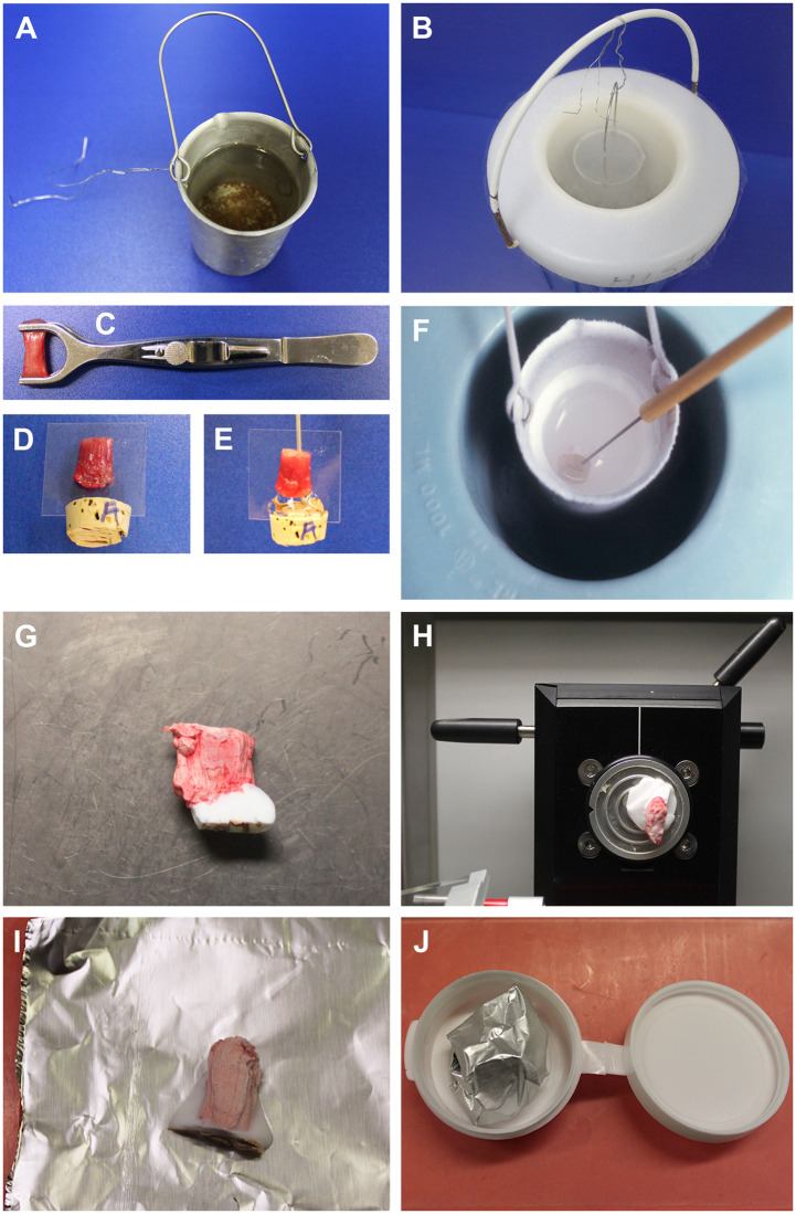
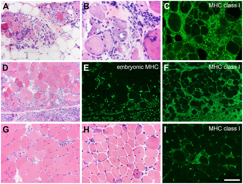
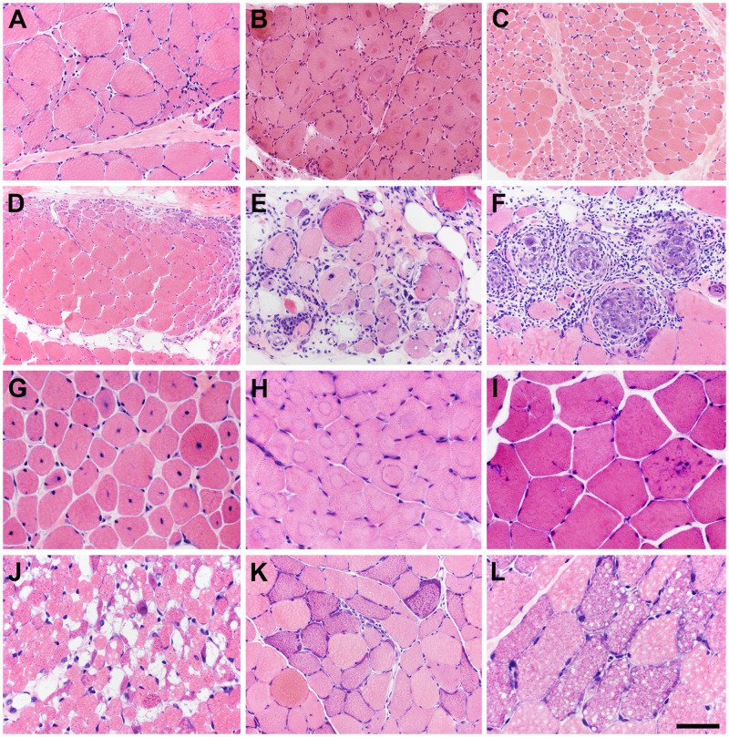
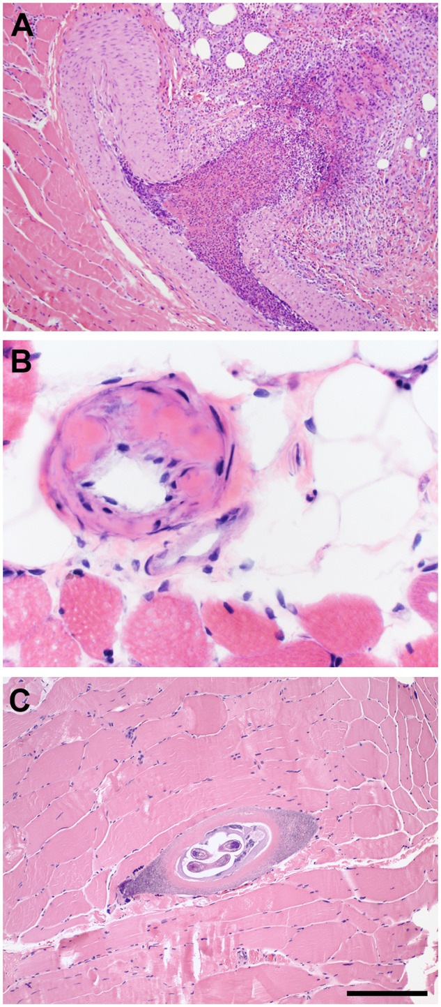
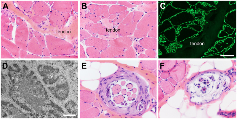
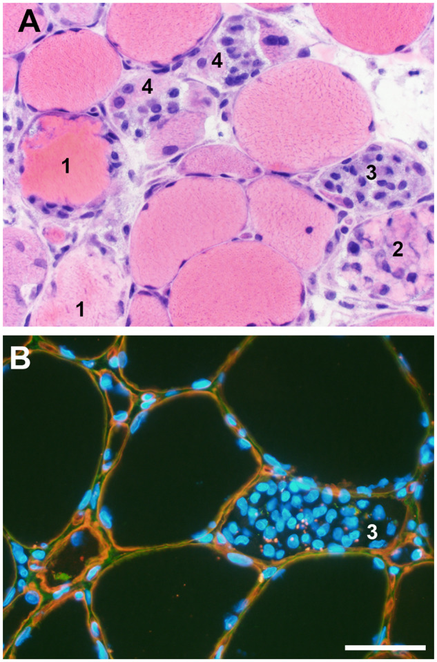
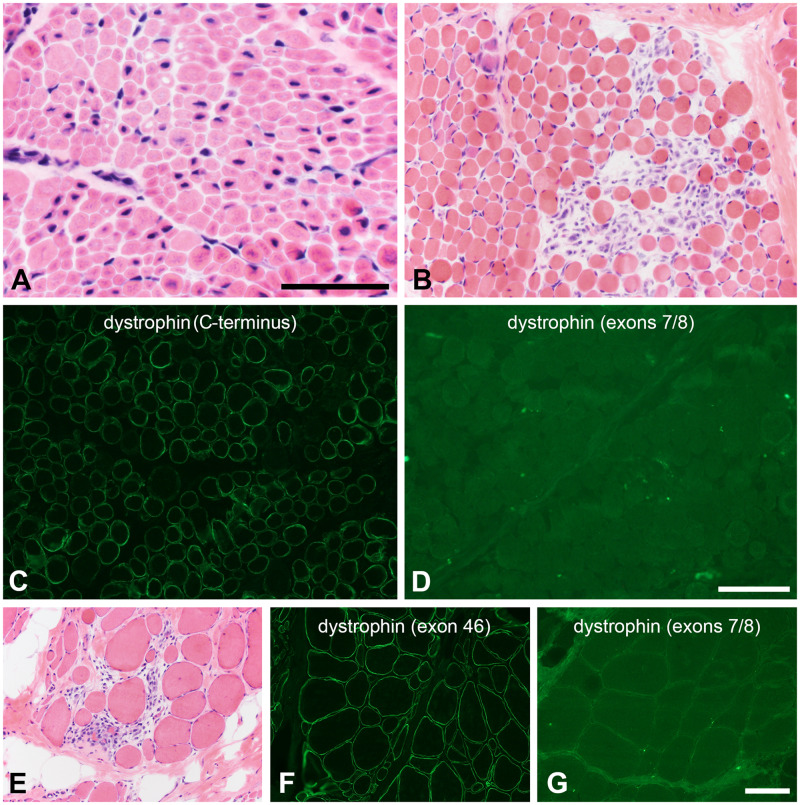
Competence in muscle biopsy evaluation is a core component of neuropathology practice. The practicing neuropathologist should be able to prepare frozen sections of muscle biopsies with minimal artifacts and identify key histopathologic features of neuromuscular disease in hematoxylin and eosin-stained sections as well as implement and interpret a basic panel of additional histochemical, enzyme histochemical, and immunohistochemical stains. Important to everyday practice is a working knowledge of normal muscle histology at different ages, muscle motor units, pitfalls of myotendinous junctions, nonpathologic variations encountered at traditional and nontraditional muscle sites, the pathophysiology of myonecrosis and regeneration, and approaches to distinguish muscular dystrophies from inflammatory myopathies and other necrotizing myopathies. Here, we provide a brief overview of what every neuropathologist needs to know concerning the muscle biopsy.
肌肉活检评估的能力是神经病理学实践的核心组成部分。执业神经病理学家应该能够用最小的伪影准备肌肉活检的冷冻切片,并在苏木精和伊红染色切片中识别神经肌肉疾病的关键组织病理学特征,以及实施和解释基本的一组额外的组织化学、酶组织化学和免疫组织化学染色。日常实践中重要的是了解不同年龄段的正常肌肉组织学、肌肉运动单位、肌腱结合处的陷阱、传统和非传统肌肉部位遇到的非病理性变化、肌肉坏死和再生的病理生理学,以及区分肌营养不良症与炎症性肌病和其他坏死性肌病的方法。在这里,我们简要概述了每位神经病理学家都需要了解的有关肌肉活检的内容。