Department of Ophthalmology, University of Muenster Medical Center, Albert-Schweitzer-Campus 1, Building D15, 48149, Muenster, Germany.
Graefes Arch Clin Exp Ophthalmol. 2020 Oct;258(10):2263-2269. doi: 10.1007/s00417-020-04788-4. Epub 2020 Jun 12.
To evaluate the retinal microvascular density using optical coherence tomography angiography (OCTA) in patients with systemic lupus erythematosus (SLE) treated with hydroxychloroquine (HCQ).
Nineteen eyes of 19 patients with SLE (study group) without HCQ retinopathy and 19 eyes of 19 healthy subjects (control group) were included in this study. The study group was divided into patients using HCQ for > 5 years (high-risk group) and < 5 years (low-risk group). The VD data of the 3 × 3 mm OCT angiogram of the superficial capillary plexus (SCP) and the choriocapillaris (VD-CC), the foveal avascular zone (FAZ) area and the central retinal thickness (CRT) were extracted and analyzed.
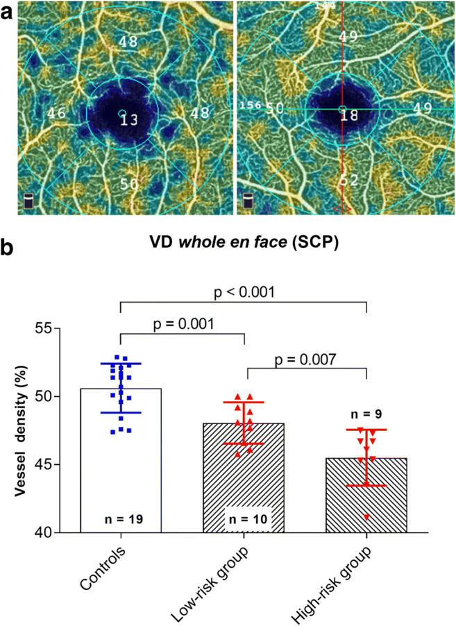
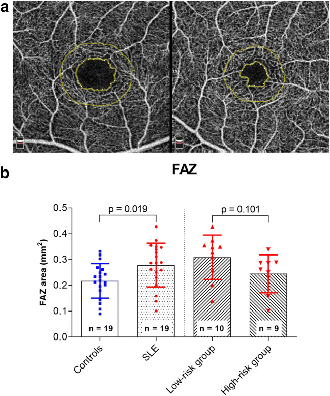
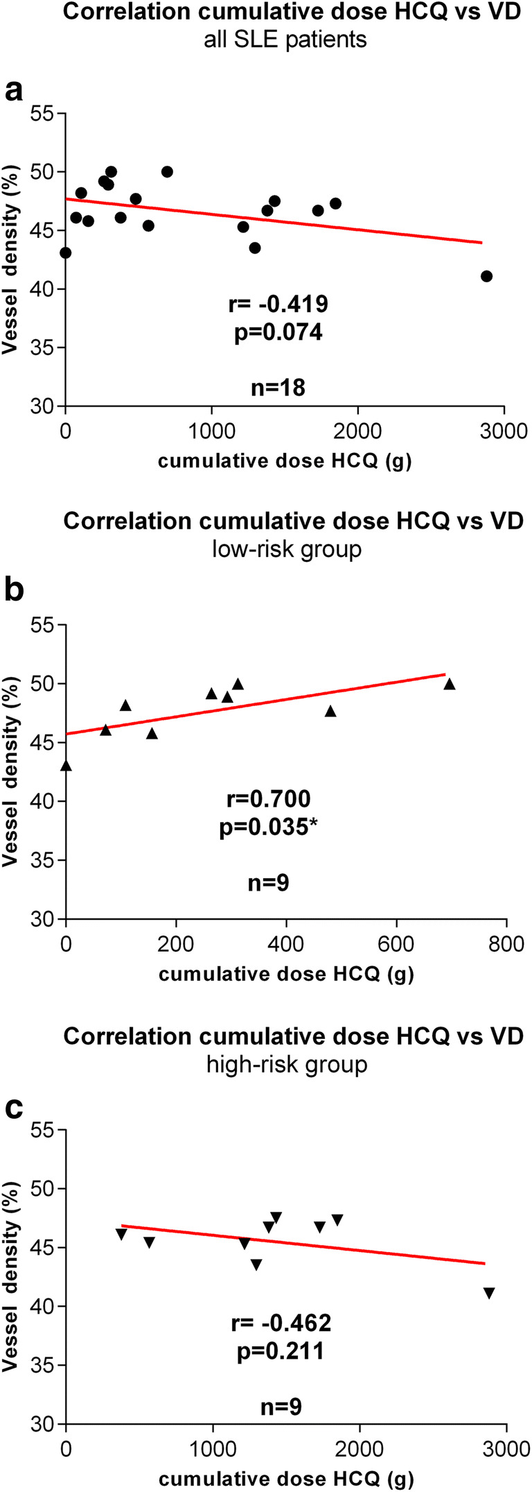
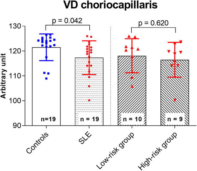
VD in the en face SCP was significantly reduced in the high-risk group and the low-risk group compared with that in the control group (p < 0.001, p = 0.001) and in the high-risk group compared with the low-risk group (p = 0.007). Correlation analysis between the cumulative dose of HCQ and the VD of the study group revealed a negative correlation, but no statistical significance (p = 0.074). However, a significant positive correlation was observed for the low-risk group (p = 0.035). In patients with SLE, VD-CC was lower (p = 0.042) and the FAZ area larger (p = 0.019). CRT showed no difference between the groups (p = 0.183).
In this study, SLE patients showed a reduced VD in both groups. In patients treated with HCQ < 5 years, HCQ might have a protective effect on retinal microvasculature. Analysis of retinal microvascular density using OCTA could be useful in the diagnosis and monitoring of vascular alteration in patients with SLE.
利用光相干断层扫描血管造影术(OCTA)评估系统性红斑狼疮(SLE)患者接受羟氯喹(HCQ)治疗后的视网膜微血管密度。
本研究纳入了 19 例 SLE 患者(研究组)的 19 只眼,这些患者均无 HCQ 视网膜病变且未使用 HCQ 治疗;同时纳入了 19 名健康受试者的 19 只眼作为对照组。研究组根据 HCQ 使用时间(> 5 年或< 5 年)分为高危组和低危组。提取并分析 3×3mm OCTA 浅层毛细血管丛(SCP)和脉络膜毛细血管(VD-CC)的血管密度(VD)、黄斑无血管区(FAZ)面积和中心视网膜厚度(CRT)数据。
与对照组相比,高危组和低危组的 SCP 面平均血管密度显著降低(p<0.001,p=0.001),且高危组显著低于低危组(p=0.007)。HCQ 累积剂量与研究组 VD 之间的相关性分析显示存在负相关,但无统计学意义(p=0.074)。然而,低危组显示出显著的正相关(p=0.035)。SLE 患者脉络膜毛细血管密度(VD-CC)较低(p=0.042),FAZ 面积较大(p=0.019)。两组 CRT 无差异(p=0.183)。
本研究中,两组 SLE 患者的 VD 均降低。在接受 HCQ 治疗< 5 年的患者中,HCQ 可能对视网膜微血管具有保护作用。OCTA 分析视网膜微血管密度可能有助于诊断和监测 SLE 患者的血管改变。