• 文献检索
  • 文档翻译
  • 深度研究
  • 学术资讯
  • Suppr Zotero 插件Zotero 插件
  • 邀请有礼
  • 套餐&价格
  • 历史记录
应用&插件
Suppr Zotero 插件Zotero 插件浏览器插件Mac 客户端Windows 客户端微信小程序
定价
高级版会员购买积分包购买API积分包
服务
文献检索文档翻译深度研究API 文档MCP 服务
关于我们
关于 Suppr公司介绍联系我们用户协议隐私条款
关注我们

Suppr 超能文献

核心技术专利:CN118964589B侵权必究
粤ICP备2023148730 号-1Suppr @ 2026

文献检索

告别复杂PubMed语法,用中文像聊天一样搜索,搜遍4000万医学文献。AI智能推荐,让科研检索更轻松。

立即免费搜索

文件翻译

保留排版,准确专业,支持PDF/Word/PPT等文件格式,支持 12+语言互译。

免费翻译文档

深度研究

AI帮你快速写综述,25分钟生成高质量综述,智能提取关键信息,辅助科研写作。

立即免费体验

正畸治疗中的脱粘与清洁:不同技术及牙釉质表面微观形态学方面的评估

Debonding and Clean-Up in Orthodontics: Evaluation of Different Techniques and Micro-Morphological Aspects of the Enamel Surface.

作者信息

D'Amario Maurizio, Bernardi Sara, Di Lauro Daniele, Marzo Giuseppe, Macchiarelli Guido, Capogreco Mario

机构信息

Department of Life, Health and Environmental Sciences, University of L'Aquila, 67100 L'Aquila, Italy.

Microscopy Center, University of L'Aquila, 67100 L'Aquila, Italy.

出版信息

Dent J (Basel). 2020 Jun 17;8(2):58. doi: 10.3390/dj8020058.

DOI:10.3390/dj8020058
PMID:32560482
原文链接:https://pmc.ncbi.nlm.nih.gov/articles/PMC7344892/
Abstract

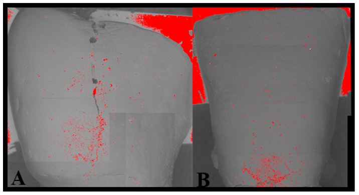
There is currently no consensus on the best way to remove adhesive remnants from teeth following debonding. The main objective of this study is to evaluate and compare the effectiveness of four adhesive resin removal (clean-up) techniques, performed with or without the use of an operative microscope. Forty human teeth were duplicated using an epoxy resin for impregnation. Brackets were bonded to teeth and debonded from teeth. Then, the samples were randomly divided into two equal groups-the naked eye group and the magnification group-and further subdivided into four equal subgroups, in order to compare the different techniques used for the clean-up. Each subgroup was formed of five natural teeth with the respective pre- and post-bonding replicas. Macro- and micro-analysis by means of a stereomicroscope and scanning electron microscopy evaluated, qualitatively and quantitatively, the adhesive remnant index and the damage index of the enamel. Overall, the magnification improved the removal of resins compared to the naked eye ( < 0.001), and the use of magnification constantly reduced resin residual and surface damage. Enamel damage and adhesive residual from the clean-up procedures represent an ascertained risk in orthodontics. The use of a magnification system improves the quality of debonding and clean-up techniques in a significant way.

摘要

目前,对于牙齿脱粘后去除粘结剂残留的最佳方法尚无共识。本研究的主要目的是评估和比较四种粘结树脂去除(清理)技术在使用或不使用手术显微镜情况下的有效性。使用环氧树脂浸渍法复制了40颗人牙。将托槽粘结到牙齿上,然后再从牙齿上取下。接着,将样本随机分为两组,每组20个,分别为肉眼组和放大组,再将每组进一步细分为四个亚组,每组5个,以比较不同的清理技术。每个亚组由五颗天然牙及其粘结前后的复制牙组成。通过体视显微镜和扫描电子显微镜进行宏观和微观分析,对粘结剂残留指数和牙釉质损伤指数进行了定性和定量评估。总体而言,与肉眼相比,放大操作改善了树脂的去除效果(P<0.001),并且使用放大设备持续减少了树脂残留和表面损伤。正畸治疗中,清理过程造成的牙釉质损伤和粘结剂残留是确定存在的风险。使用放大系统能显著提高脱粘和清理技术的质量。

https://cdn.ncbi.nlm.nih.gov/pmc/blobs/8a35/7344892/5eb96e9986d6/dentistry-08-00058-g004.jpg

相似文献

1
Debonding and Clean-Up in Orthodontics: Evaluation of Different Techniques and Micro-Morphological Aspects of the Enamel Surface.正畸治疗中的脱粘与清洁:不同技术及牙釉质表面微观形态学方面的评估
Dent J (Basel). 2020 Jun 17;8(2):58. doi: 10.3390/dj8020058.
2
A Comparative Evaluation of Enamel Surface Roughness of Two Different Bonding Adhesives After Debonding With Atomic Force Microscopy.使用原子力显微镜对两种不同粘结剂脱粘后牙釉质表面粗糙度的比较评估
Cureus. 2022 Nov 18;14(11):e31661. doi: 10.7759/cureus.31661. eCollection 2022 Nov.
3
Evaluation of enamel surface integrity after orthodontic bracket debonding: comparison of three different system.正畸托槽去粘接后牙釉质表面完整性的评价:三种不同系统的比较。
BMC Oral Health. 2024 Mar 20;24(1):358. doi: 10.1186/s12903-024-04138-4.
4
Enamel evaluation by scanning electron microscopy after debonding brackets and removal of adhesive remnants.在去除托槽并清除粘结剂残余物后,通过扫描电子显微镜对牙釉质进行评估。
J Clin Exp Dent. 2018 Mar 1;10(3):e248-e251. doi: 10.4317/jced.54553. eCollection 2018 Mar.
5
Effects of various debonding and adhesive clearance methods on enamel surface: an in vitro study.不同脱粘和粘结剂清除方法对牙釉质表面的影响:一项体外研究。
BMC Oral Health. 2017 Feb 27;17(1):58. doi: 10.1186/s12903-017-0349-6.
6
Comparison of the debonding characteristics of two innovative ceramic bracket designs.两种新型陶瓷托槽设计的脱粘特性比较。
Am J Orthod Dentofacial Orthop. 1999 Jul;116(1):86-92. doi: 10.1016/s0889-5406(99)70307-0.
7
Effect of orthodontic debonding and residual adhesive removal on 3D enamel microroughness.正畸去粘结及去除残留粘结剂对三维釉质微粗糙度的影响。
PeerJ. 2016 Oct 11;4:e2558. doi: 10.7717/peerj.2558. eCollection 2016.
8
Evaluation of enamel roughness after orthodontic debonding and clean-up procedures using zirconia, tungsten carbide, and white stone burs: an in vitro study.评价氧化锆、碳化钨和白宝石车针对正畸去带及清洁后牙釉质粗糙度的影响:一项体外研究。
BMC Oral Health. 2023 Jul 13;23(1):478. doi: 10.1186/s12903-023-03194-6.
9
Ceramic bracket debonding: the evaluation of two debonding techniques and their effect on enamel.陶瓷托槽拆除:两种拆除技术的评估及其对牙釉质的影响。
Aust Orthod J. 1994 Mar;13(2):80-5.
10
Comparison of Enamel Surface Roughness after Bracket Debonding and Adhesive Resin Removal Using Different Burs with and without the Aid of a Magnifying Loupe.比较使用带/不带放大镜助视器的不同车针在托槽去除和粘结剂去除后对釉质表面粗糙度的影响。
J Contemp Dent Pract. 2022 Nov 1;23(11):1091-1099. doi: 10.5005/jp-journals-10024-3432.

引用本文的文献

1
Evaluation of Enamel Surface Roughness and Volumetric Change after Resin Remnant Removal following Orthodontic Bracket Debonding.正畸托槽脱粘后去除树脂残余物后牙釉质表面粗糙度和体积变化的评估。
Oral Health Prev Dent. 2025 Jul 4;23:355-364. doi: 10.3290/j.ohpd.c_2117.
2
Magnification: The game changer in dentistry.放大技术:牙科领域的变革者。
World J Methodol. 2025 Jun 20;15(2):100937. doi: 10.5662/wjm.v15.i2.100937.
3
Functional modification and antibacterial evaluation of orthodontic adhesives with poly (lysine)-derived carbon dots.

本文引用的文献

1
Commercial oral hygiene products and implant collar surfaces: Scanning electron microscopy observations.商用口腔卫生产品和种植体颈部表面:扫描电子显微镜观察。
Can J Dent Hyg. 2020 Feb 1;54(1):26-31.
2
Microbiological and SEM-EDS Evaluation of Titanium Surfaces Exposed to Periodontal Gel: In Vitro Study.暴露于牙周凝胶的钛表面的微生物学和扫描电子显微镜-能谱分析评估:体外研究
Materials (Basel). 2019 May 4;12(9):1448. doi: 10.3390/ma12091448.
3
The importance of Post Mortem Computed Tomography (PMCT) in the reconstruction of the bullet trajectory.
聚(赖氨酸)衍生碳点对正畸粘合剂的功能改性及抗菌性能评价
RSC Adv. 2025 Feb 20;15(8):5876-5888. doi: 10.1039/d4ra08710k. eCollection 2025 Feb 19.
4
Benefits of using magnification in dental specialties - a narrative review.牙科专业中使用放大技术的益处——一篇叙述性综述。
Med Pharm Rep. 2023 Jul;96(3):254-257. doi: 10.15386/mpr-2556. Epub 2023 Jul 27.
5
Bond Strength Survival of a Novel Calcium Phosphate-Enriched Orthodontic Self-Etching System after Various Ageing Protocols: An In Vitro Study.不同老化方案后新型富磷酸钙正畸自酸蚀系统的粘结强度及存活率:一项体外研究
Int J Dent. 2022 Dec 31;2022:3960362. doi: 10.1155/2022/3960362. eCollection 2022.
6
Fluorescence-Aided Identification Technique (FIT) Improves Tooth Surface Clean-Up after Debonding of Buccal and Lingual Orthodontic Appliances.荧光辅助识别技术(FIT)改善颊侧和舌侧正畸矫治器脱粘后牙面清洁效果。
J Clin Med. 2021 Dec 31;11(1):213. doi: 10.3390/jcm11010213.
7
Vertical and Orovestibular Forces Generated by Beta-Titanium and Stainless-Steel Rectangular Wires in Labial and Fully Customized Lingual Bracket Systems.β钛合金和不锈钢矩形丝在唇侧及全定制舌侧托槽系统中产生的垂直和口前庭力
Materials (Basel). 2021 Sep 28;14(19):5632. doi: 10.3390/ma14195632.
8
Efficient Design of a Clear Aligner Attachment to Induce Bodily Tooth Movement in Orthodontic Treatment Using Finite Element Analysis.使用有限元分析进行正畸治疗中诱导牙齿整体移动的透明矫治器附件的高效设计
Materials (Basel). 2021 Aug 30;14(17):4926. doi: 10.3390/ma14174926.
9
Effect of Polishing Systems on the Surface Roughness of Nano-Hybrid and Nano-Filling Composite Resins: A Systematic Review.抛光系统对纳米混合和纳米填充复合树脂表面粗糙度的影响:一项系统评价。
Dent J (Basel). 2021 Aug 12;9(8):95. doi: 10.3390/dj9080095.
10
Comparative Analysis of Stress in the Periodontal Ligament and Center of Rotation in the Tooth after Orthodontic Treatment Depending on Clear Aligner Thickness-Finite Element Analysis Study.基于隐形矫治器厚度的正畸治疗后牙周膜应力与牙齿旋转中心的对比分析——有限元分析研究
Materials (Basel). 2021 Jan 9;14(2):324. doi: 10.3390/ma14020324.
尸检计算机断层扫描(PMCT)在子弹弹道重建中的重要性。
Clin Ter. 2019 Mar-Apr;170(2):e129-e133. doi: 10.7417/CT.2019.2122.
4
The Two by Four appliance: a nationwide cross-sectional survey.两板四钉器具:全国性横断面调查。
Eur J Paediatr Dent. 2018 Jun;19(2):145-150. doi: 10.23804/ejpd.2018.19.02.09.
5
Scanning electron microscopy and microbiological approaches for the evaluation of salivary microorganisms behaviour on anatase titanium surfaces: In vitro study.扫描电子显微镜和微生物学方法用于评估锐钛矿型钛表面唾液微生物行为:体外研究
Morphologie. 2018 Mar;102(336):1-6. doi: 10.1016/j.morpho.2017.12.001. Epub 2017 Dec 26.
6
Quantitative analysis of enamel on debonded orthodontic brackets.正畸托槽脱粘后釉质的定量分析。
Am J Orthod Dentofacial Orthop. 2017 Sep;152(3):312-319. doi: 10.1016/j.ajodo.2017.01.020.
7
Histomorphometric Parameters of the Growth Plate and Trabecular Bone in Wild-Type and Trefoil Factor Family 3 (Tff3)-Deficient Mice Analyzed by Free and Open-Source Image Processing Software.利用免费开源图像处理软件分析野生型和三叶因子家族3(Tff3)基因缺陷小鼠生长板和小梁骨的组织形态计量学参数
Microsc Microanal. 2017 Aug;23(4):818-825. doi: 10.1017/S1431927617000630.
8
Comparative study of dental enamel loss after debonding braces by analytical scanning electron microscopy (SEM).通过分析扫描电子显微镜(SEM)对去除牙套后牙釉质损失的对比研究。
Microsc Res Tech. 2017 Jul;80(7):680-686. doi: 10.1002/jemt.22849. Epub 2017 Feb 5.
9
Efficacy of auxiliary devices for removal of fluorescent residue after bracket debonding.正畸托槽拆除后用于去除荧光残留辅助装置的疗效
Angle Orthod. 2017 May;87(3):440-447. doi: 10.2319/081916-629.1. Epub 2016 Dec 30.
10
In vivo confocal microscopy analysis of enamel defects after orthodontic treatment: A preliminary study.正畸治疗后牙釉质缺陷的体内共聚焦显微镜分析:一项初步研究。
Ultrastruct Pathol. 2016 Nov-Dec;40(6):317-323. doi: 10.1080/01913123.2016.1237603. Epub 2016 Oct 13.