Department of Diagnosis and Surgery, Sao Paulo State University - Unesp. School of Dentistry, Rua José Bonifácio, 1193, Araçatuba, ZIP code:, CEP16015-050, Sao Paulo, Brazil.
Undergradutate student, Sao Paulo State University - Unesp. School of Dentistry, Rua José Bonifácio, 1193, Araçatuba, ZIP code:, CEP16015-050, Sao Paulo, Brazil.
Sci Rep. 2020 Jun 19;10(1):10000. doi: 10.1038/s41598-020-65289-2.
In this in vivo animal study, we evaluated the effect of plasma electrolytic oxidation (PEO) coating on the topographic and biological parameters of implants installed in rats with induced osteoporosis and low-quality bones.
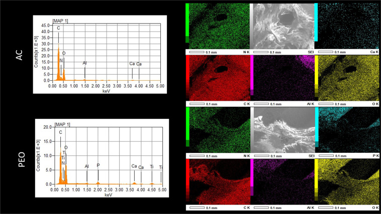
In total 44 Wistar rats (Rattus novergicus), 6 months old, were submitted to ovariectomy (OXV group) and dummy surgery (SHAM group). After 90 days, the ELISA test was performed and the ovariectomy effectiveness was confirmed. In each tibial metaphysis, an implant with PEO coating containing Ca and P molecules were installed, and the other tibia received an implant with SLA acid etching and blasting (AC) (control surface). After 42 days, 16 rats from each group were euthanized, their tibias were removed for histological and immunohistochemical analysis (OPG, RANKL, OC and TRAP), as well as reverse torque biomechanics. Data were submitted to One-way ANOVA or Kruskal-Wallis tests, followed by a Tukey post-test; P < 0.05. Histological analyses showed higher bone neoformation values among the members of the PEO group, SHAM and OVX groups. Immunohistochemical analysis demonstrated equilibrium in all groups when comparing surfaces for TRAP, OC and RANKL (P > 0.05), whereas OPG showed higher PEO labeling in the OVX group (P < 0.05). Biomechanical analysis showed higher reverse torque values (N.cm) for PEO, irrespective of whether they were OVX or SHAM groups (P < 0.05).
The results indicated that the PEO texturing method favored bone formation and showed higher bone maturation levels during later periods in osteoporotic rats.
在这项体内动物研究中,我们评估了等离子电解氧化(PEO)涂层对植入患有骨质疏松症和低质量骨骼的大鼠的种植体的形貌和生物学参数的影响。
共有 44 只 6 个月大的 Wistar 大鼠(Rattus novergicus)接受卵巢切除术(OVX 组)和假手术(SHAM 组)。90 天后,进行 ELISA 测试以确认卵巢切除术的效果。在每个胫骨干骺端安装含有 Ca 和 P 分子的 PEO 涂层植入物,而另一个胫骨则接受 SLA 酸蚀和喷砂(AC)(对照表面)植入物。42 天后,每组处死 16 只大鼠,取出胫骨进行组织学和免疫组织化学分析(OPG、RANKL、OC 和 TRAP)以及反向扭矩生物力学分析。数据采用单因素方差分析或 Kruskal-Wallis 检验,然后进行 Tukey 事后检验;P<0.05。组织学分析显示,PEO 组、SHAM 组和 OVX 组的成员的新骨形成值更高。免疫组织化学分析表明,在比较 TRAP、OC 和 RANKL 表面时,所有组之间均处于平衡状态(P>0.05),而 OPG 在 OVX 组中显示出更高的 PEO 标记(P<0.05)。生物力学分析显示,PEO 组的反向扭矩值(N.cm)更高,无论它们是 OVX 组还是 SHAM 组(P<0.05)。
结果表明,PEO 纹理化方法有利于骨形成,并在骨质疏松大鼠的后期表现出更高的骨成熟水平。