Almuhayawi Mohammed S, Ramadan Wafaa S, Harakeh Steve, Al Jaouni Soad K, Bharali Dhruba J, Mousa Shaker A, Almuhayawi Saad M
Department of Medical Microbiology and Parasitology, Faculty of Medicine, King Abdulaziz University (KAU), Jeddah, Saudi Arabia.
Department of Anatomy, Faculty of Medicine (FM), KAU, Saudi Arabia.
Saudi J Biol Sci. 2020 Jul;27(7):1710-1716. doi: 10.1016/j.sjbs.2020.04.045. Epub 2020 May 7.
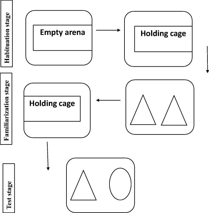
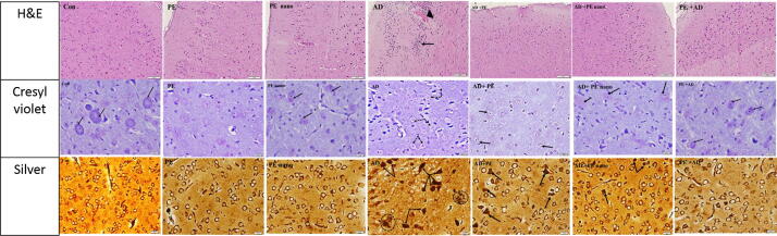
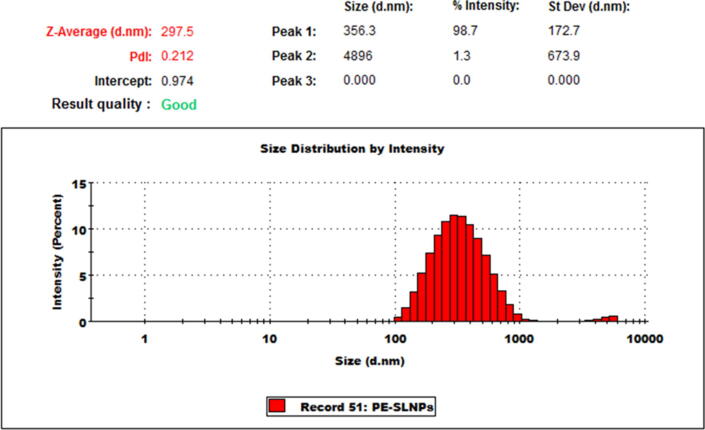
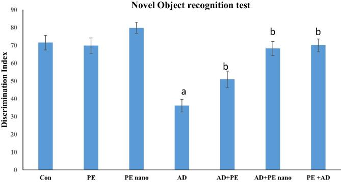
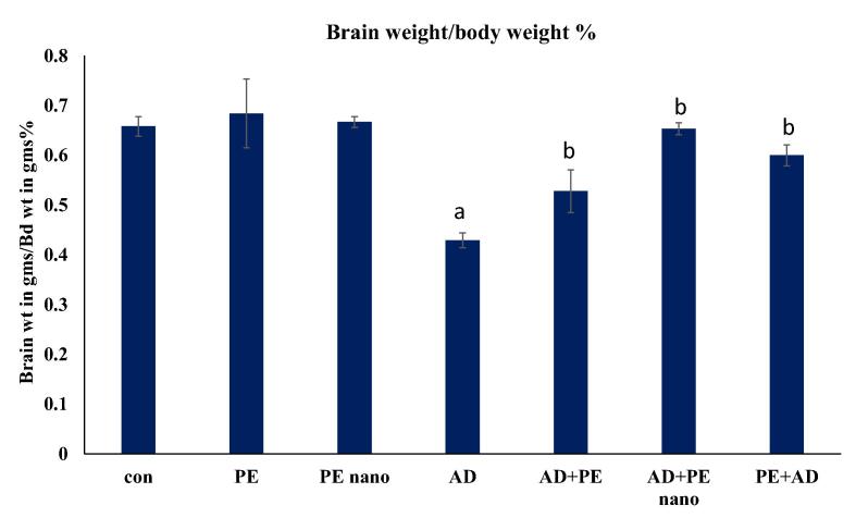
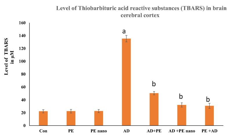
The oxidative stress leading to degenerative changes in the brain of Alzheimer's disease (AD) is evident. Our aim was to evaluate the therapeutic and protective effects of pomegranate extract (PE) and pomegranate extract-loaded nanoparticles (PE nano) in an AlCl 3-induced AD rat model. Nanoparticles were synthesized with a PE load of 0.68% w/w, and 70 male Wistar rats were divided into 7 groups: Group I was the control, Group II received PE., Group III received PE nano for 2 weeks, Group IV received AlCl 3 (50 mg/kg) daily orally for 4 weeks, Group V received PE for 2 weeks, Group VI received PE nano for 2 weeks, and Groups V and VI were started after AlCl 3 administration was stopped. Group VII received PE for 2 weeks and was stopped before AlCl 3 was administered. The Results revealed that the discrimination index in the novel object recognition test was the least in AD rat model but increased in cases protected with PE treated with PE nano. Similar results were shown based on calculating the brain weight/body weight percent. The biomarkers of antioxidant activity (catalase, glutathione and total antioxidant activity) in brain homogenate were significantly increased in groups treated with either PE or PE nano. The thiobarbituric acid reactive substance measured to estimate lipid peroxidation was significantly increased in AD rat model and decreased in groups protected with PE or treated with PE nano. Histopathological studies using hematoxylin and eosin, cresyl violet, and silver stains revealed hyaline degeneration, chromatolysis, and hallmarks of AD; neurofibrillary tangles and the senile plaques in brains of AD rat model. Restoration of the histological architecture, Nissl granules, and minimal appearance of hallmarks of AD characterized brains treated with PE or PE nano. In conclusion, PE was more effective as a protectant than a therapeutic measure in alleviating the antioxidant, lipid peroxidative effects and histopathological hallmarks in AD brains. But, the therapeutic PE-loaded nanoparticles increased the efficacy of active components and produced similar results as the protective PE.
导致阿尔茨海默病(AD)大脑退行性变化的氧化应激是明显的。我们的目的是评估石榴提取物(PE)和负载石榴提取物的纳米颗粒(PE纳米颗粒)在氯化铝诱导的AD大鼠模型中的治疗和保护作用。合成了负载量为0.68%(w/w)的纳米颗粒,将70只雄性Wistar大鼠分为7组:第一组为对照组,第二组接受PE,第三组接受PE纳米颗粒2周,第四组每天口服氯化铝(50mg/kg)4周,第五组接受PE 2周,第六组接受PE纳米颗粒2周,第五组和第六组在停止给予氯化铝后开始给药。第七组接受PE 2周,并在给予氯化铝之前停药。结果显示,在新物体识别试验中,AD大鼠模型的辨别指数最低,但在用PE或PE纳米颗粒处理的受保护病例中有所增加。根据计算脑重/体重百分比也显示了类似结果。在用PE或PE纳米颗粒处理的组中,脑匀浆中抗氧化活性的生物标志物(过氧化氢酶、谷胱甘肽和总抗氧化活性)显著增加。为评估脂质过氧化而测量的硫代巴比妥酸反应性物质在AD大鼠模型中显著增加,而在用PE或PE纳米颗粒处理的组中则降低。使用苏木精和伊红、甲酚紫和银染进行的组织病理学研究显示,AD大鼠模型的大脑存在透明变性、染色质溶解以及AD的特征性表现;神经原纤维缠结和老年斑。用PE或PE纳米颗粒处理的大脑的组织学结构、尼氏体颗粒得以恢复,AD特征性表现最少。总之,在减轻AD大脑中的抗氧化、脂质过氧化作用和组织病理学特征方面,PE作为一种保护剂比治疗措施更有效。但是,治疗性的负载PE的纳米颗粒提高了活性成分的疗效,并产生了与保护性PE相似的结果。