• 文献检索
  • 文档翻译
  • 深度研究
  • 学术资讯
  • Suppr Zotero 插件Zotero 插件
  • 邀请有礼
  • 套餐&价格
  • 历史记录
应用&插件
Suppr Zotero 插件Zotero 插件浏览器插件Mac 客户端Windows 客户端微信小程序
定价
高级版会员购买积分包购买API积分包
服务
文献检索文档翻译深度研究API 文档MCP 服务
关于我们
关于 Suppr公司介绍联系我们用户协议隐私条款
关注我们

Suppr 超能文献

核心技术专利:CN118964589B侵权必究
粤ICP备2023148730 号-1Suppr @ 2026

文献检索

告别复杂PubMed语法,用中文像聊天一样搜索,搜遍4000万医学文献。AI智能推荐,让科研检索更轻松。

立即免费搜索

文件翻译

保留排版,准确专业,支持PDF/Word/PPT等文件格式,支持 12+语言互译。

免费翻译文档

深度研究

AI帮你快速写综述,25分钟生成高质量综述,智能提取关键信息,辅助科研写作。

立即免费体验

颅内粥样硬化斑块的定量直方图分析:一项高分辨率磁共振成像研究。

Quantitative Histogram Analysis on Intracranial Atherosclerotic Plaques: A High-Resolution Magnetic Resonance Imaging Study.

机构信息

Department of Radiology (Z.S., J. Li, W.P., T.J., Q.L., J. Lu), Changhai Hospital, Naval Medical University, Shanghai, China.

Department of Radiology, University of Cambridge, United Kingdom (Z.S., Z.M., Z.T.).

出版信息

Stroke. 2020 Jul;51(7):2161-2169. doi: 10.1161/STROKEAHA.120.029062. Epub 2020 Jun 17.

DOI:10.1161/STROKEAHA.120.029062
PMID:32568660
原文链接:https://pmc.ncbi.nlm.nih.gov/articles/PMC7306260/
Abstract

BACKGROUND AND PURPOSE

Intracranial atherosclerosis is one of the main causes of stroke, and high-resolution magnetic resonance imaging provides useful imaging biomarkers related to the risk of ischemic events. This study aims to evaluate differences in histogram features between culprit and nonculprit intracranial atherosclerosis using high-resolution magnetic resonance imaging.

METHODS

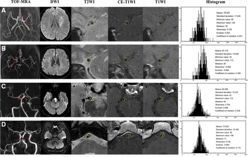
Two hundred forty-seven patients with intracranial atherosclerosis who underwent high-resolution magnetic resonance imaging sequentially between January 2015 and December 2016 were recruited. Quantitative features, including stenosis, plaque burden, minimum luminal area, intraplaque hemorrhage, enhancement ratio, and dispersion of signal intensity (coefficient of variation), were analyzed based on T2-, T1-, and contrast-enhanced T1-weighted images. Step-wise regression analysis was used to identify key determinates differentiating culprit and nonculprit plaques and to calculate the odds ratios (ORs) with 95% CIs.

RESULTS

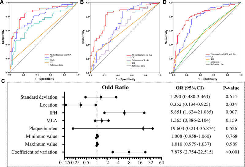
In total, 190 plaques were identified, of which 88 plaques (37 culprit and 51 nonculprit) were located in the middle cerebral artery and 102 (57 culprit and 45 nonculprit) in the basilar artery. Nearly 90% of culprit lesions had a degree of luminal stenosis of <70%. Multiple logistic regression analyses showed that intraplaque hemorrhage (OR, 16.294 [95% CI, 1.043-254.632]; =0.047), minimum luminal area (OR, 1.468 [95% CI, 1.032-2.087]; =0.033), and coefficient of variation (OR, 13.425 [95% CI, 3.987-45.204]; <0.001) were 3 significant features in defining culprit plaques in middle cerebral artery. The enhancement ratio (OR, 9.476 [95% CI, 1.256-71.464]; =0.029), intraplaque hemorrhage (OR, 2.847 [95% CI, 0.971-10.203]; =0.046), and coefficient of variation (OR, 10.068 [95% CI, 2.820-21.343]; <0.001) were significantly associated with plaque type in basilar artery. Coefficient of variation was a strong independent predictor in defining plaque type for both middle cerebral artery and basilar artery with sensitivity, specificity, and accuracy being 0.79, 0.80, and 0.80, respectively.

CONCLUSIONS

Features characterized by high-resolution magnetic resonance imaging provided complementary values over luminal stenosis in defined lesion type for intracranial atherosclerosis; the dispersion of signal intensity in histogram analysis was a particularly effective predictive parameter.

摘要

背景与目的

颅内动脉粥样硬化是中风的主要原因之一,高分辨率磁共振成像提供了与缺血事件风险相关的有用成像生物标志物。本研究旨在使用高分辨率磁共振成像评估罪魁祸首和非罪魁祸首颅内动脉粥样硬化斑块之间的直方图特征差异。

方法

2015 年 1 月至 2016 年 12 月期间,连续招募了 247 例接受高分辨率磁共振成像的颅内动脉粥样硬化患者。基于 T2、T1 和对比增强 T1 加权图像,分析了定量特征,包括狭窄程度、斑块负荷、最小管腔面积、斑块内出血、增强比和信号强度分布(变异系数)。采用逐步回归分析来确定区分罪魁祸首和非罪魁祸首斑块的关键决定因素,并计算 95%置信区间的优势比(OR)。

结果

共发现 190 个斑块,其中 88 个斑块(37 个罪魁祸首和 51 个非罪魁祸首)位于大脑中动脉,102 个斑块(57 个罪魁祸首和 45 个非罪魁祸首)位于基底动脉。近 90%的罪魁祸首病变的管腔狭窄程度<70%。多因素逻辑回归分析显示,斑块内出血(OR,16.294 [95%CI,1.043-254.632];=0.047)、最小管腔面积(OR,1.468 [95%CI,1.032-2.087];=0.033)和变异系数(OR,13.425 [95%CI,3.987-45.204];<0.001)是确定大脑中动脉罪魁祸首斑块的 3 个重要特征。增强比(OR,9.476 [95%CI,1.256-71.464];=0.029)、斑块内出血(OR,2.847 [95%CI,0.971-10.203];=0.046)和变异系数(OR,10.068 [95%CI,2.820-21.343];<0.001)与基底动脉斑块类型显著相关。变异系数是定义大脑中动脉和基底动脉斑块类型的独立强预测因子,其敏感性、特异性和准确性分别为 0.79、0.80 和 0.80。

结论

高分辨率磁共振成像特征在定义颅内动脉粥样硬化病变类型方面提供了比管腔狭窄更具互补价值的信息;直方图分析中的信号强度分布是一种特别有效的预测参数。

https://cdn.ncbi.nlm.nih.gov/pmc/blobs/ce4f/7306260/64e8d71cf61d/str-51-2161-g009.jpg
https://cdn.ncbi.nlm.nih.gov/pmc/blobs/ce4f/7306260/2b5943949b97/str-51-2161-g005.jpg
https://cdn.ncbi.nlm.nih.gov/pmc/blobs/ce4f/7306260/64e8d71cf61d/str-51-2161-g009.jpg
https://cdn.ncbi.nlm.nih.gov/pmc/blobs/ce4f/7306260/2b5943949b97/str-51-2161-g005.jpg
https://cdn.ncbi.nlm.nih.gov/pmc/blobs/ce4f/7306260/64e8d71cf61d/str-51-2161-g009.jpg

相似文献

1
Quantitative Histogram Analysis on Intracranial Atherosclerotic Plaques: A High-Resolution Magnetic Resonance Imaging Study.颅内粥样硬化斑块的定量直方图分析:一项高分辨率磁共振成像研究。
Stroke. 2020 Jul;51(7):2161-2169. doi: 10.1161/STROKEAHA.120.029062. Epub 2020 Jun 17.
2
Characteristics of culprit intracranial plaque without substantial stenosis in ischemic stroke using three-dimensional high-resolution vessel wall magnetic resonance imaging.使用三维高分辨率血管壁磁共振成像对缺血性卒中中无显著狭窄的罪犯颅内斑块的特征分析
Front Neurosci. 2023 Mar 23;17:1160018. doi: 10.3389/fnins.2023.1160018. eCollection 2023.
3
Gadolinium enhancement of atherosclerotic plaque in the intracranial artery.颅内动脉粥样硬化斑块的钆增强。
Neurol Res. 2021 Dec;43(12):1040-1049. doi: 10.1080/01616412.2021.1949682. Epub 2021 Jul 6.
4
Detection and Quantification of Symptomatic Atherosclerotic Plaques With High-Resolution Imaging in Cryptogenic Stroke.高分辨率成像技术在不明原因卒中患者中检测和定量分析症状性动脉粥样硬化斑块
Stroke. 2020 Dec;51(12):3623-3631. doi: 10.1161/STROKEAHA.120.031167. Epub 2020 Oct 1.
5
Association of Hypertension With Both Occurrence and Outcome of Symptomatic Patients With Mild Intracranial Atherosclerotic Stenosis: A Prospective Higher Resolution Magnetic Resonance Imaging Study.高血压与症状性轻度颅内动脉粥样硬化狭窄患者的发生和结局的相关性:一项前瞻性高分辨率磁共振成像研究。
J Magn Reson Imaging. 2021 Jul;54(1):76-88. doi: 10.1002/jmri.27516. Epub 2021 Mar 10.
6
An assessment on the incremental value of high-resolution magnetic resonance imaging to identify culprit plaques in atherosclerotic disease of the middle cerebral artery.关于高分辨率磁共振成像在识别大脑中动脉动脉粥样硬化疾病罪犯斑块方面的增量价值评估。
Eur Radiol. 2016 Jul;26(7):2206-14. doi: 10.1007/s00330-015-4008-5. Epub 2015 Sep 16.
7
Plaque Distribution and Characteristics in Low-Grade Middle Cerebral Artery Stenosis and Its Clinical Relevance: A 3-Dimensional High-Resolution Magnetic Resonance Imaging Study.大脑中动脉低度狭窄的斑块分布、特征及其临床相关性:一项三维高分辨率磁共振成像研究
J Stroke Cerebrovasc Dis. 2018 Aug;27(8):2243-2249. doi: 10.1016/j.jstrokecerebrovasdis.2018.04.010. Epub 2018 May 8.
8
Identification of high-risk plaque features in intracranial atherosclerosis: initial experience using a radiomic approach.颅内动脉粥样硬化斑块高危特征的识别:一种基于放射组学方法的初步经验。
Eur Radiol. 2018 Sep;28(9):3912-3921. doi: 10.1007/s00330-018-5395-1. Epub 2018 Apr 9.
9
Co-existing intracranial and extracranial carotid artery atherosclerotic plaques and recurrent stroke risk: a three-dimensional multicontrast cardiovascular magnetic resonance study.颅内和颅外颈动脉粥样硬化斑块并存与复发性卒中风险:一项三维多对比心血管磁共振研究
J Cardiovasc Magn Reson. 2016 Dec 2;18(1):90. doi: 10.1186/s12968-016-0309-3.
10
Follow-up assessment of atherosclerotic plaques in acute ischemic stroke patients using high-resolution vessel wall MR imaging.采用高分辨率血管壁磁共振成像对急性缺血性脑卒中患者的动脉粥样硬化斑块进行随访评估。
Neuroradiology. 2022 Dec;64(12):2257-2266. doi: 10.1007/s00234-022-03002-y. Epub 2022 Jun 29.

引用本文的文献

1
Inter-rater reliability of atherosclerotic plaque enhancement on 7 T vessel wall MRI: Comparison with quantitative analysis.7T血管壁磁共振成像中动脉粥样硬化斑块强化的评分者间可靠性:与定量分析的比较
Interv Neuroradiol. 2025 Aug 24:15910199251368719. doi: 10.1177/15910199251368719.
2
Endothelial Glycocalyx in Cerebral Infarction After Endovascular Treatment in Patients With Intracranial Artery Stenosis.颅内动脉狭窄患者血管内治疗后脑梗死中的内皮糖萼
CNS Neurosci Ther. 2025 Aug;31(8):e70545. doi: 10.1111/cns.70545.
3
Quantitative plaque characteristics based on 3D high-resolution vessel wall imaging predict long-term recurrence in intracranial atherosclerotic stroke.

本文引用的文献

1
The utility of MRI histogram and texture analysis for the prediction of histological diagnosis in head and neck malignancies.MRI 直方图和纹理分析在头颈部恶性肿瘤组织学诊断预测中的应用。
Cancer Imaging. 2019 Feb 4;19(1):5. doi: 10.1186/s40644-019-0193-9.
2
Clinical Significance of Intraplaque Hemorrhage in Low- and High-Grade Basilar Artery Stenosis on High-Resolution MRI.高分辨率 MRI 显示低级别和高级别基底动脉狭窄斑块内出血的临床意义。
AJNR Am J Neuroradiol. 2018 Jul;39(7):1286-1292. doi: 10.3174/ajnr.A5676. Epub 2018 May 24.
3
Identification of high-risk plaque features in intracranial atherosclerosis: initial experience using a radiomic approach.
基于三维高分辨率血管壁成像的定量斑块特征可预测颅内动脉粥样硬化性卒中的长期复发。
Neuroradiology. 2025 Jun 19. doi: 10.1007/s00234-025-03642-w.
4
High-risk characteristics of recurrent ischemic stroke after intensive medical management for 6-month follow-up: a histogram study on vessel wall MRI.强化内科治疗6个月随访后复发性缺血性卒中的高危特征:基于血管壁MRI的直方图研究
Eur Radiol. 2025 Mar;35(3):1313-1324. doi: 10.1007/s00330-024-11304-3. Epub 2025 Jan 2.
5
Comparison of computer-aided quantitative measurement and physician visual assessment in the evaluation of intracranial atherosclerotic stenosis: a vessel wall magnetic resonance imaging study.计算机辅助定量测量与医生视觉评估在颅内动脉粥样硬化狭窄评估中的比较:一项血管壁磁共振成像研究
Quant Imaging Med Surg. 2024 Oct 1;14(10):7459-7471. doi: 10.21037/qims-24-788. Epub 2024 Sep 26.
6
7T-high resolution MRI-derived radiomic analysis for the identification of symptomatic intracranial atherosclerotic plaques.用于识别有症状颅内动脉粥样硬化斑块的7T高分辨率MRI衍生的放射组学分析
Interv Neuroradiol. 2024 Aug 30:15910199241275722. doi: 10.1177/15910199241275722.
7
Deep learning improves quality of intracranial vessel wall MRI for better characterization of potentially culprit plaques.深度学习提高颅内血管壁 MRI 质量,更好地对潜在罪犯斑块进行特征描述。
Sci Rep. 2024 Aug 16;14(1):18983. doi: 10.1038/s41598-024-69750-4.
8
The hemodynamic and geometric characteristics of carotid artery atherosclerotic plaque formation.颈动脉粥样硬化斑块形成的血流动力学和几何特征。
Quant Imaging Med Surg. 2024 Jul 1;14(7):4348-4361. doi: 10.21037/qims-23-1827. Epub 2024 Jun 11.
9
Differential diagnosis of thyroid nodules using heterogeneity quantification software on ultrasound images: correlation with the Bethesda system and surgical pathology.超声图像异质性量化软件在甲状腺结节鉴别诊断中的应用:与 Bethesda 系统及外科病理学的相关性。
Sci Rep. 2024 May 4;14(1):10288. doi: 10.1038/s41598-024-60881-2.
10
A clinical-radiomics combined model based on carotid atherosclerotic plaque for prediction of ischemic stroke.基于颈动脉粥样硬化斑块的临床-放射组学联合模型用于预测缺血性卒中
Front Neurol. 2024 Mar 13;15:1343423. doi: 10.3389/fneur.2024.1343423. eCollection 2024.
颅内动脉粥样硬化斑块高危特征的识别:一种基于放射组学方法的初步经验。
Eur Radiol. 2018 Sep;28(9):3912-3921. doi: 10.1007/s00330-018-5395-1. Epub 2018 Apr 9.
4
Magnetic resonance imaging of intracranial atherosclerosis: Comparison of ex vivo 3T MRI and histologic findings.颅内动脉粥样硬化的磁共振成像:离体 3T MRI 与组织学发现的比较。
Eur J Radiol. 2017 Dec;97:110-114. doi: 10.1016/j.ejrad.2017.10.013. Epub 2017 Oct 18.
5
N-(carboxymethyl)lysine Concentration in Debris from Carotid Artery Stenting Correlates Independently with Signal Intensity on T1-Weighted Black-Blood Magnetic Resonance Images.颈动脉支架置入术后碎屑中N-(羧甲基)赖氨酸浓度与T1加权黑血磁共振图像上的信号强度独立相关。
J Stroke Cerebrovasc Dis. 2017 Jun;26(6):1341-1348. doi: 10.1016/j.jstrokecerebrovasdis.2017.02.005. Epub 2017 Mar 14.
6
Histopathological Differences Between the Anterior and Posterior Brain Arteries as a Function of Aging.作为衰老函数的大脑前动脉和后动脉之间的组织病理学差异。
Stroke. 2017 Mar;48(3):638-644. doi: 10.1161/STROKEAHA.116.015630. Epub 2017 Feb 14.
7
Classification of Carotid Plaque Echogenicity by Combining Texture Features and Morphologic Characteristics.结合纹理特征和形态学特征对颈动脉斑块回声性进行分类
J Ultrasound Med. 2016 Oct;35(10):2253-61. doi: 10.7863/ultra.15.09002. Epub 2016 Aug 31.
8
Intracranial Vessel Wall MRI: Principles and Expert Consensus Recommendations of the American Society of Neuroradiology.颅内血管壁磁共振成像:美国神经放射学会的原理与专家共识推荐
AJNR Am J Neuroradiol. 2017 Feb;38(2):218-229. doi: 10.3174/ajnr.A4893. Epub 2016 Jul 28.
9
Ex-vivo imaging and plaque type classification of intracranial atherosclerotic plaque using high resolution MRI.使用高分辨率磁共振成像对颅内动脉粥样硬化斑块进行体外成像及斑块类型分类
Atherosclerosis. 2016 Jun;249:10-6. doi: 10.1016/j.atherosclerosis.2016.03.033. Epub 2016 Mar 30.
10
MR Imaging Measures of Intracranial Atherosclerosis in a Population-based Study.一项基于人群的研究中颅内动脉粥样硬化的磁共振成像测量
Radiology. 2016 Sep;280(3):860-8. doi: 10.1148/radiol.2016151124. Epub 2016 Mar 29.