• 文献检索
  • 文档翻译
  • 深度研究
  • 学术资讯
  • Suppr Zotero 插件Zotero 插件
  • 邀请有礼
  • 套餐&价格
  • 历史记录
应用&插件
Suppr Zotero 插件Zotero 插件浏览器插件Mac 客户端Windows 客户端微信小程序
定价
高级版会员购买积分包购买API积分包
服务
文献检索文档翻译深度研究API 文档MCP 服务
关于我们
关于 Suppr公司介绍联系我们用户协议隐私条款
关注我们

Suppr 超能文献

核心技术专利:CN118964589B侵权必究
粤ICP备2023148730 号-1Suppr @ 2026

文献检索

告别复杂PubMed语法,用中文像聊天一样搜索,搜遍4000万医学文献。AI智能推荐,让科研检索更轻松。

立即免费搜索

文件翻译

保留排版,准确专业,支持PDF/Word/PPT等文件格式,支持 12+语言互译。

免费翻译文档

深度研究

AI帮你快速写综述,25分钟生成高质量综述,智能提取关键信息,辅助科研写作。

立即免费体验

尺寸排阻色谱法与超速离心法相结合可改善血浆来源的小细胞外囊泡的蛋白质组分析。

Combination of Size-Exclusion Chromatography and Ultracentrifugation Improves the Proteomic Profiling of Plasma-Derived Small Extracellular Vesicles.

作者信息

Wei Rui, Zhao Libo, Kong Guanyi, Liu Xiang, Zhu Shengtao, Zhang Shutian, Min Li

机构信息

Department of Gastroenterology, Beijing Friendship Hospital, Capital Medical University, National Clinical Research Center for Digestive Disease, Beijing Digestive Disease Center, Beijing Key Laboratory for Precancerous Lesion of Digestive Disease, Beijing, 100050 P. R. China.

Echo Biotech Co., Ltd, Beijing, 100010 P. R. China.

出版信息

Biol Proced Online. 2020 Jun 23;22:12. doi: 10.1186/s12575-020-00125-5. eCollection 2020.

DOI:10.1186/s12575-020-00125-5
PMID:32587481
原文链接:https://pmc.ncbi.nlm.nih.gov/articles/PMC7313174/
Abstract

BACKGROUND

Circulating small extracellular vesicles (sEVs) and its associated proteins are of great interest in the early detection of many diseases. However, there is no gold standard for plasma sEVs isolation, especially for proteomic profiling which could be largely affected by contamination such as lipoproteins and plasma proteins. Previous studies suggested combinations of different sEVs isolation methods could improve the yield and purity of the isolated fractions. Nevertheless, there is no systematic evaluation of size-exclusion chromatography (SEC), ultracentrifugation (UC), and their combination in a proteomic perspective.

RESULTS

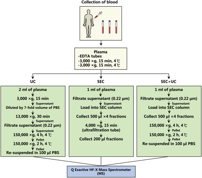
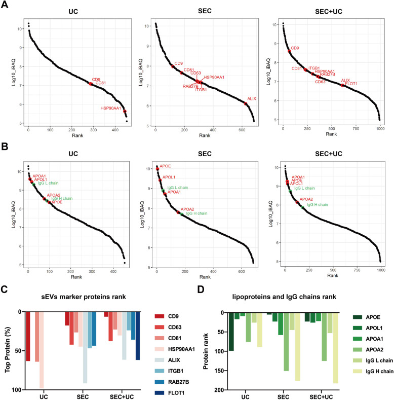
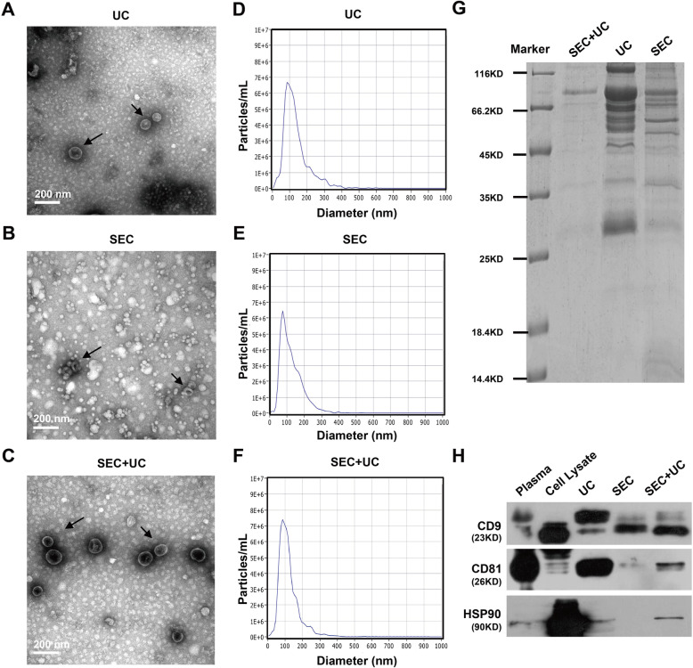
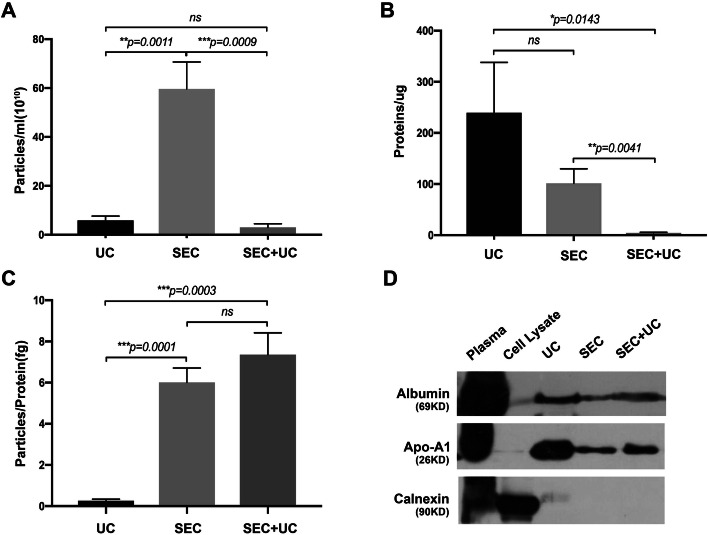
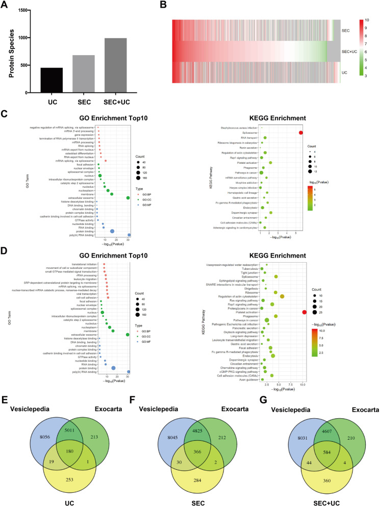
Plasma samples were collected from healthy individuals, and sEVs were separated by one-step SEC, one-step UC, and combining SEC with UC, respectively. Here we exhibited that the purity of sEVs was improved by SEC in contrast to traditional UC. Furthermore, by conducting a SEC procedure followed by UC, we separated sEVs with the highest purity. In the proteomic analysis, 992 protein species were identified in the plasma sEVs isolated by our novel separation method, of which several proteins are sEVs-associated proteins but hitherto never been identified in the previous studies and database, much more than plasma sEVs isolated by UC (453) or SEC (682) alone. As compared to Vesiclepedia and Exocarta databases, plasma sEVs isolated by the new procedure kept 584 previously identified sEVs-associated proteins and 360 other proteins that have not been detected before. Detailed analysis suggested that more kinds of sEVs biomarkers, such as CD9, ALIX, and FLOT1, could be identified in plasma sEVs isolated by the novel isolation method as compared to one-step UC/SEC. Furthermore, the lower abundance ranks of common contaminants, such as lipoproteins and IgG chains, in the sEVs fractions obtained by our new method as compared to one-step UC/SEC also demonstrated the purity of sEVs had been improved.

CONCLUSIONS

Combining SEC with UC could significantly improve the performance of mass spectrometry-based proteomic profiling in analyzing plasma-derived sEVs.

摘要

背景

循环小细胞外囊泡(sEVs)及其相关蛋白在多种疾病的早期检测中备受关注。然而,血浆sEVs分离尚无金标准,尤其是蛋白质组分析,其可能在很大程度上受到脂蛋白和血浆蛋白等污染的影响。先前的研究表明,不同sEVs分离方法的组合可提高分离组分的产量和纯度。然而,从蛋白质组学角度对尺寸排阻色谱法(SEC)、超速离心法(UC)及其组合尚无系统评估。

结果

收集健康个体的血浆样本,分别通过一步SEC、一步UC以及SEC与UC相结合的方法分离sEVs。我们在此表明,与传统UC相比,SEC提高了sEVs的纯度。此外,通过先进行SEC程序再进行UC,我们分离出了纯度最高的sEVs。在蛋白质组分析中,通过我们的新型分离方法分离的血浆sEVs中鉴定出992种蛋白质,其中几种蛋白质是sEVs相关蛋白,但此前在以往研究和数据库中从未被鉴定出来,这比单独通过UC(453种)或SEC(682种)分离的血浆sEVs要多得多。与Vesiclepedia和Exocarta数据库相比,通过新程序分离的血浆sEVs保留了584种先前鉴定的sEVs相关蛋白和360种以前未检测到的其他蛋白。详细分析表明,与一步UC/SEC相比,通过新型分离方法分离的血浆sEVs中可鉴定出更多种类的sEVs生物标志物,如CD9、ALIX和FLOT1。此外,与一步UC/SEC相比,我们新方法获得的sEVs组分中常见污染物(如脂蛋白和IgG链)的丰度排名更低,这也证明了sEVs的纯度得到了提高。

结论

将SEC与UC相结合可显著提高基于质谱的蛋白质组分析在分析血浆来源sEVs中的性能。

https://cdn.ncbi.nlm.nih.gov/pmc/blobs/44ce/7313174/fe62672554a1/12575_2020_125_Fig5_HTML.jpg
https://cdn.ncbi.nlm.nih.gov/pmc/blobs/44ce/7313174/013e3938d69b/12575_2020_125_Fig1_HTML.jpg
https://cdn.ncbi.nlm.nih.gov/pmc/blobs/44ce/7313174/6571d4d0d250/12575_2020_125_Fig2_HTML.jpg
https://cdn.ncbi.nlm.nih.gov/pmc/blobs/44ce/7313174/d4c71747137b/12575_2020_125_Fig3_HTML.jpg
https://cdn.ncbi.nlm.nih.gov/pmc/blobs/44ce/7313174/2893a4c0531b/12575_2020_125_Fig4_HTML.jpg
https://cdn.ncbi.nlm.nih.gov/pmc/blobs/44ce/7313174/fe62672554a1/12575_2020_125_Fig5_HTML.jpg

相似文献

1
Combination of Size-Exclusion Chromatography and Ultracentrifugation Improves the Proteomic Profiling of Plasma-Derived Small Extracellular Vesicles.尺寸排阻色谱法与超速离心法相结合可改善血浆来源的小细胞外囊泡的蛋白质组分析。
Biol Proced Online. 2020 Jun 23;22:12. doi: 10.1186/s12575-020-00125-5. eCollection 2020.
2
Comparison of small extracellular vesicles isolated from plasma by ultracentrifugation or size-exclusion chromatography: yield, purity and functional potential.通过超速离心或尺寸排阻色谱法从血浆中分离的小细胞外囊泡的比较:产量、纯度和功能潜力。
J Extracell Vesicles. 2018 Dec 28;8(1):1560809. doi: 10.1080/20013078.2018.1560809. eCollection 2019.
3
A Comparison of Blood Plasma Small Extracellular Vesicle Enrichment Strategies for Proteomic Analysis.用于蛋白质组学分析的血浆小细胞外囊泡富集策略比较
Proteomes. 2022 Jun 1;10(2):19. doi: 10.3390/proteomes10020019.
4
Comparison of proteomic landscape of extracellular vesicles in pleural effusions isolated by three strategies.三种策略分离的胸腔积液中细胞外囊泡蛋白质组景观的比较
Front Bioeng Biotechnol. 2023 Apr 12;11:1108952. doi: 10.3389/fbioe.2023.1108952. eCollection 2023.
5
Isolating Small Extracellular Vesicles from Small Volumes of Blood Plasma using size exclusion chromatography and density gradient ultracentrifugation: A Comparative Study.使用尺寸排阻色谱法和密度梯度超速离心法从少量血浆中分离小细胞外囊泡:一项比较研究。
bioRxiv. 2025 Jan 22:2023.10.30.564707. doi: 10.1101/2023.10.30.564707.
6
Optimal Isolation Method of Small Extracellular Vesicles from Rat Plasma.从大鼠血浆中分离小细胞外囊泡的最佳方法。
Int J Mol Sci. 2019 Sep 26;20(19):4780. doi: 10.3390/ijms20194780.
7
Coupling size exclusion chromatography to ultracentrifugation improves detection of exosomal proteins from human plasma by LC-MS.将尺寸排阻色谱与超速离心相结合,可通过液相色谱-质谱联用(LC-MS)提高从人血浆中检测外泌体蛋白的能力。
Pract Lab Med. 2021 Jun 23;26:e00241. doi: 10.1016/j.plabm.2021.e00241. eCollection 2021 Aug.
8
An Isolation System to Collect High Quality and Purity Extracellular Vesicles from Serum.一种从血清中收集高质量和高纯度细胞外囊泡的分离系统。
Int J Nanomedicine. 2021 Sep 29;16:6681-6692. doi: 10.2147/IJN.S328325. eCollection 2021.
9
Comparing the Proteomic Profiles of Extracellular Vesicles Isolated using Different Methods from Long-term Stored Plasma Samples.比较从长期储存的血浆样本中使用不同方法分离得到的细胞外囊泡的蛋白质组学图谱。
Biol Proced Online. 2024 Jun 19;26(1):18. doi: 10.1186/s12575-024-00243-4.
10
Improving the Purity of Extracellular Vesicles by Removal of Lipoproteins from Size Exclusion Chromatography- and Ultracentrifugation-Processed Samples Using Glycosaminoglycan-Functionalized Magnetic Beads.使用糖胺聚糖功能化的磁性珠从分子筛层析和超速离心处理的样品中去除脂蛋白来提高细胞外囊泡的纯度。
ACS Appl Mater Interfaces. 2024 Aug 28;16(34):44386-44398. doi: 10.1021/acsami.4c03869. Epub 2024 Aug 16.

引用本文的文献

1
MicroRNA-6084 orchestrates angiogenesis and liver metastasis in colorectal cancer via extracellular vesicles.微小RNA-6084通过细胞外囊泡调控结直肠癌的血管生成和肝转移。
JCI Insight. 2025 Jun 10;10(14). doi: 10.1172/jci.insight.189503. eCollection 2025 Jul 22.
2
Carotid Plaque-Derived Small Extracellular Vesicles Mediate Atherosclerosis and Correlate With Plaque Vulnerability.颈动脉斑块来源的小细胞外囊泡介导动脉粥样硬化并与斑块易损性相关。
MedComm (2020). 2025 May 19;6(6):e70220. doi: 10.1002/mco2.70220. eCollection 2025 Jun.
3
Development of LncRNA Biomarkers in Extracellular Vesicle of Amniotic Fluid Associated with Antenatal Hydronephrosis.

本文引用的文献

1
Quality and efficiency assessment of six extracellular vesicle isolation methods by nano-flow cytometry.通过纳米流式细胞术对六种细胞外囊泡分离方法的质量和效率评估
J Extracell Vesicles. 2019 Nov 29;9(1):1697028. doi: 10.1080/20013078.2019.1697028. eCollection 2020.
2
An Update on Isolation Methods for Proteomic Studies of Extracellular Vesicles in Biofluids.生物体液中外泌体蛋白质组学研究的分离方法更新。
Molecules. 2019 Sep 27;24(19):3516. doi: 10.3390/molecules24193516.
3
Evaluation of circulating small extracellular vesicles derived miRNAs as biomarkers of early colon cancer: a comparison with plasma total miRNAs.
与产前肾积水相关的羊水细胞外囊泡中长链非编码RNA生物标志物的研究进展
Biomedicines. 2025 Mar 8;13(3):668. doi: 10.3390/biomedicines13030668.
4
Plasma extracellular vesicles from recurrent GBMs carrying LDHA to activate glioblastoma stemness by enhancing glycolysis.来自复发性胶质母细胞瘤的血浆细胞外囊泡携带乳酸脱氢酶A,通过增强糖酵解来激活胶质母细胞瘤干性。
Theranostics. 2025 Feb 26;15(8):3655-3672. doi: 10.7150/thno.102014. eCollection 2025.
5
The proteomic landscape of stool-derived extracellular vesicles in patients with pre-cancerous lesions and colorectal cancer.癌前病变和结直肠癌患者粪便来源细胞外囊泡的蛋白质组学图谱。
Commun Biol. 2025 Feb 13;8(1):228. doi: 10.1038/s42003-025-07652-5.
6
Plasma-derived extracellular vesicles prime alveolar macrophages for autophagy and ferroptosis in sepsis-induced acute lung injury.血浆来源的细胞外囊泡使肺泡巨噬细胞对脓毒症诱导的急性肺损伤中的自噬和铁死亡产生预适应。
Mol Med. 2025 Feb 4;31(1):40. doi: 10.1186/s10020-025-01111-x.
7
Unveiling the Emerging Role of Extracellular Vesicle-Inflammasomes in Hyperoxia-Induced Neonatal Lung and Brain Injury.揭示细胞外囊泡炎性小体在高氧诱导的新生儿肺和脑损伤中的新作用。
Cells. 2024 Dec 18;13(24):2094. doi: 10.3390/cells13242094.
8
Extracellular vesicles as a promising biomarker resource in liquid biopsy for cancer.细胞外囊泡作为液体活检中一种很有前景的癌症生物标志物来源。
Extracell Vesicles Circ Nucl Acids. 2021 May 13;2(2):148-174. doi: 10.20517/evcna.2021.06. eCollection 2021.
9
Revisiting the diagnostic performance of exosomes: harnessing the feasibility of combinatorial exosomal miRNA profiles for colorectal cancer diagnosis.重新审视外泌体的诊断性能:利用组合外泌体微小RNA谱进行结直肠癌诊断的可行性。
Discov Oncol. 2024 Oct 30;15(1):605. doi: 10.1007/s12672-024-01481-4.
10
Proteomic and metabolomic profiles of plasma-derived Extracellular Vesicles differentiate melanoma patients from healthy controls.血浆来源的细胞外囊泡的蛋白质组学和代谢组学图谱可区分黑色素瘤患者与健康对照。
Transl Oncol. 2024 Dec;50:102152. doi: 10.1016/j.tranon.2024.102152. Epub 2024 Oct 13.
评估循环小细胞外囊泡衍生的miRNA作为早期结肠癌生物标志物:与血浆总miRNA的比较
J Extracell Vesicles. 2019 Jul 22;8(1):1643670. doi: 10.1080/20013078.2019.1643670. eCollection 2019.
4
Reassessment of Exosome Composition.重新评估外泌体组成。
Cell. 2019 Apr 4;177(2):428-445.e18. doi: 10.1016/j.cell.2019.02.029.
5
Extracellular vesicle isolation methods: rising impact of size-exclusion chromatography.细胞外囊泡分离方法:尺寸排阻色谱法的影响日益增大。
Cell Mol Life Sci. 2019 Jun;76(12):2369-2382. doi: 10.1007/s00018-019-03071-y. Epub 2019 Mar 19.
6
Comparison of small extracellular vesicles isolated from plasma by ultracentrifugation or size-exclusion chromatography: yield, purity and functional potential.通过超速离心或尺寸排阻色谱法从血浆中分离的小细胞外囊泡的比较:产量、纯度和功能潜力。
J Extracell Vesicles. 2018 Dec 28;8(1):1560809. doi: 10.1080/20013078.2018.1560809. eCollection 2019.
7
Minimal information for studies of extracellular vesicles 2018 (MISEV2018): a position statement of the International Society for Extracellular Vesicles and update of the MISEV2014 guidelines.细胞外囊泡研究的最低限度信息2018(MISEV2018):国际细胞外囊泡协会的立场声明及MISEV2014指南的更新
J Extracell Vesicles. 2018 Nov 23;7(1):1535750. doi: 10.1080/20013078.2018.1535750. eCollection 2018.
8
Balancing yield, purity and practicality: a modified differential ultracentrifugation protocol for efficient isolation of small extracellular vesicles from human serum.平衡产量、纯度和实用性:改良的差速超速离心法从人血清中高效分离小细胞外囊泡的方案。
RNA Biol. 2019 Jan;16(1):5-12. doi: 10.1080/15476286.2018.1564465. Epub 2019 Jan 13.
9
Oncogenic Regulation of Extracellular Vesicle Proteome and Heterogeneity.致癌基因对细胞外囊泡蛋白质组和异质性的调控。
Proteomics. 2019 Jan;19(1-2):e1800169. doi: 10.1002/pmic.201800169. Epub 2019 Jan 4.
10
Detailed analysis of the plasma extracellular vesicle proteome after separation from lipoproteins.详细分析脂蛋白分离后的血浆细胞外囊泡蛋白质组。
Cell Mol Life Sci. 2018 Aug;75(15):2873-2886. doi: 10.1007/s00018-018-2773-4. Epub 2018 Feb 13.