Alameldin Sara, Costina Victor, Abdel-Baset Hesham A, Nitschke Katja, Nuhn Phillip, Neumaier Michael, Hedtke Maren
Institute for Clinical Chemistry, Medical Faculty Mannheim of the University of Heidelberg, University Hospital Mannheim, Mannheim, Germany.
Clinical Pathology Department, Assiut University Hospital, Assiut, Egypt.
Pract Lab Med. 2021 Jun 23;26:e00241. doi: 10.1016/j.plabm.2021.e00241. eCollection 2021 Aug.
Exosomes are small lipid bilayer vesicles that are defined by their endocytic origin and size range of 30-140 nm. They are constantly produced by different cell types, by both healthy and abnormal cells, and can be isolated from almost all body fluids.Little information exists in isolating exosomes from plasma due to the complexity of its content and the presence of contaminating plasma proteins.
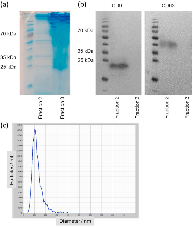
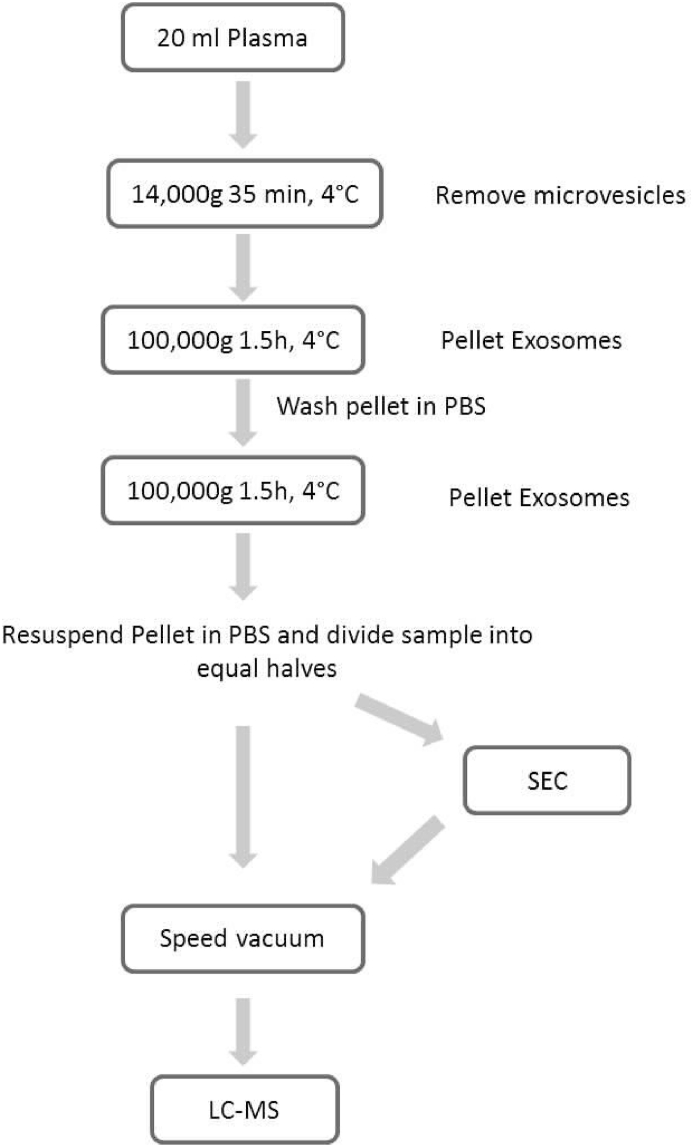
We carried-out liquid chromatography-mass spectrometry (LC-MS/MS) analyses of plasma-derived vesicles from 4 healthy donors obtained by 2 coupled methodologies: Ultracentrifugation (UC) coupled with size-exclusion chromatography (SEC) to isolate and subsequently enrich exosomes.We compared the proteins detected by UC alone and UC coupled with SEC.
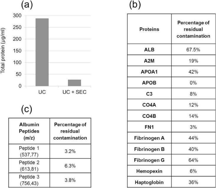
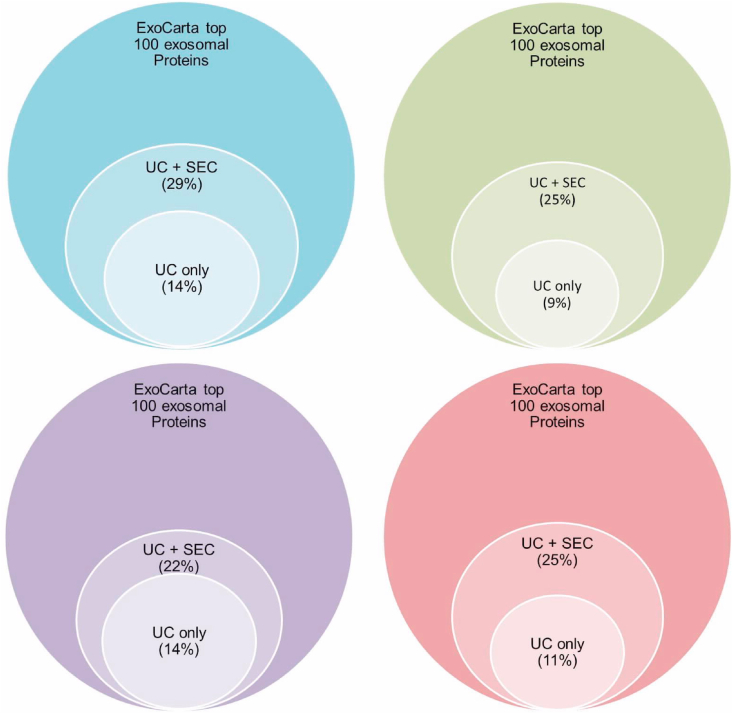
In the coupled UC + SEC methodology we found 52.25% more proteins enriched in exosomes as CD9, Annexins, YWHAZ (14-3-3 family) and others, than by using UC alone. There is also a reduction of 98.8% of contaminating plasma proteins by coupling UC and SEC in comparison to using UC alone.
We conclude that exosomes can be successfully isolated from plasma using a very simple combination of standard methods, which could largely improve the proteomics profiling of plasma exosomes.
外泌体是由其胞吞起源和30 - 140纳米大小范围所定义的小脂质双层囊泡。它们由不同细胞类型持续产生,包括健康细胞和异常细胞,并且几乎可以从所有体液中分离出来。由于血浆成分复杂且存在污染性血浆蛋白,从血浆中分离外泌体的信息较少。
我们对4名健康供体的血浆来源囊泡进行了液相色谱 - 质谱联用(LC - MS/MS)分析,通过两种联用方法获得血浆来源囊泡:超速离心(UC)结合尺寸排阻色谱(SEC)来分离并随后富集外泌体。我们比较了单独使用UC和UC结合SEC检测到的蛋白质。
在联用UC + SEC方法中,我们发现与单独使用UC相比,外泌体中富集的蛋白质如CD9、膜联蛋白、YWHAZ(14 - 3 - 3家族)等增加了52.25%。与单独使用UC相比,联用UC和SEC还使污染性血浆蛋白减少了98.8%。
我们得出结论,使用非常简单的标准方法组合可以成功从血浆中分离外泌体,这可以极大地改善血浆外泌体的蛋白质组学分析。