• 文献检索
  • 文档翻译
  • 深度研究
  • 学术资讯
  • Suppr Zotero 插件Zotero 插件
  • 邀请有礼
  • 套餐&价格
  • 历史记录
应用&插件
Suppr Zotero 插件Zotero 插件浏览器插件Mac 客户端Windows 客户端微信小程序
定价
高级版会员购买积分包购买API积分包
服务
文献检索文档翻译深度研究API 文档MCP 服务
关于我们
关于 Suppr公司介绍联系我们用户协议隐私条款
关注我们

Suppr 超能文献

核心技术专利:CN118964589B侵权必究
粤ICP备2023148730 号-1Suppr @ 2026

文献检索

告别复杂PubMed语法,用中文像聊天一样搜索,搜遍4000万医学文献。AI智能推荐,让科研检索更轻松。

立即免费搜索

文件翻译

保留排版,准确专业,支持PDF/Word/PPT等文件格式,支持 12+语言互译。

免费翻译文档

深度研究

AI帮你快速写综述,25分钟生成高质量综述,智能提取关键信息,辅助科研写作。

立即免费体验

首次逆转录聚合酶链反应(RT-PCR)检测结果为阴性的COVID-19患者的胸部CT:与首次RT-PCR检测结果为阳性的患者比较。

Chest CT of COVID-19 in patients with a negative first RT-PCR test: Comparison with patients with a positive first RT-PCR test.

作者信息

Chen Zu-Hua, Li Yun-Jiang, Wang Xiu-Juan, Ye Yun-Feng, Wu Bao-Liang, Zhang Yan, Xuan Wei-Ling, Bao Jian-Feng, Deng Xue-Ying

机构信息

Department of Radiology.

Department of Science and Education, The Hangzhou Xixi Hospital Affiliated to Zhejiang Chinese Medical University.

出版信息

Medicine (Baltimore). 2020 Jun 26;99(26):e20837. doi: 10.1097/MD.0000000000020837.

DOI:10.1097/MD.0000000000020837
PMID:32590775
原文链接:https://pmc.ncbi.nlm.nih.gov/articles/PMC7328934/
Abstract

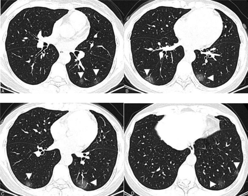
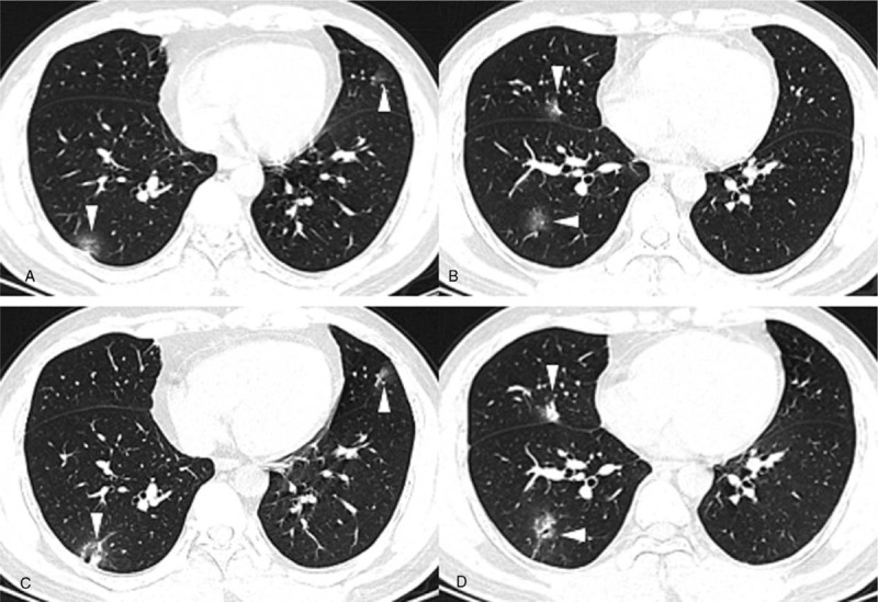
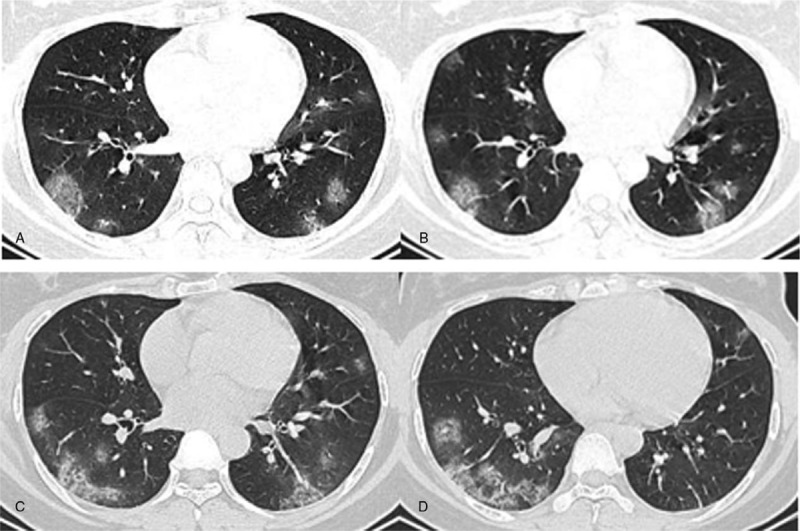
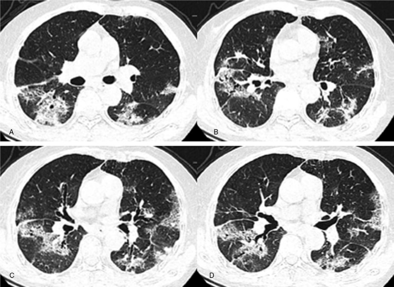
To compare clinical and imaging features between patients with an initial negative reverse-transcription-polymerase chain-reaction (RT-PCR) test and patients with an initial positive RT-PCR test. CT follow-up analysis in the negative RT-PCR group is also described.Thirty-three patients with SARS-CoV-2 infection confirmed by RT-PCR, with 216 lesions upon CT, were included. Demographic information and chest CT imaging features were collected.The average age in the whole study group was 46.9 ± 11.1 years, with 18 males and 15 females. Patients in the positive RT-PCR test group were more likely to have a fever than patients in the negative RT-PCR test group (85.7% vs 50%, P < .05). Lesions in the positive group were more likely to be located in the peripheral area than lesions in the negative group (83.6% vs 68.2%, P < .05). Regarding the appearance of 216 lesions, ground-glass opacities (GGOs) with consolidation (43.2%) was the most common appearance in the negative group, followed by pure GGOs (31.8%), while in the positive group, pure GGOs (32%) and GGOs with interlobular septal thickening (32.8%) were both most frequent, and the difference between them was evident (P < .05). For the follow-up analysis, the largest short-axis of a lesion was smaller upon follow-up (median size 13.6 mm vs 14 mm), albeit by a smaller margin. Pure GGOs decreased in frequency, from 31.3% to 21.3%, while consolidation increased in frequency, from 7.5% to 12.5%.The manifestations of COVID-19 in patients with a first negative RT-PCR test and patients with a positive first RT-PCR test are different to some extent. The consolidation component may increase after follow-up.

摘要

比较初次逆转录聚合酶链反应(RT-PCR)检测结果为阴性的患者与初次RT-PCR检测结果为阳性的患者之间的临床和影像学特征。还描述了RT-PCR检测结果为阴性的患者的CT随访分析。纳入了33例经RT-PCR确诊的新型冠状病毒肺炎(SARS-CoV-2)感染患者,其CT上共有216个病灶。收集了人口统计学信息和胸部CT影像特征。整个研究组的平均年龄为46.9±11.1岁,男性18例,女性15例。RT-PCR检测结果为阳性的患者比RT-PCR检测结果为阴性的患者更易出现发热(85.7%对50%,P<0.05)。阳性组的病灶比阴性组的病灶更易位于外周区域(83.6%对68.2%,P<0.05)。关于216个病灶的表现,在阴性组中,磨玻璃影(GGO)合并实变(43.2%)是最常见的表现,其次是单纯GGO(31.8%),而在阳性组中,单纯GGO(32%)和伴有小叶间隔增厚的GGO(32.8%)最为常见,两者之间差异明显(P<0.05)。对于随访分析,病灶的最大短轴在随访时变小(中位大小13.6mm对14mm),尽管幅度较小。单纯GGO的频率从31.3%降至21.3%,而实变的频率从7.5%增至12.5%。初次RT-PCR检测结果为阴性的患者和初次RT-PCR检测结果为阳性的患者的新型冠状病毒肺炎表现存在一定差异。随访后实变成分可能增加。

https://cdn.ncbi.nlm.nih.gov/pmc/blobs/58a3/7328934/3ab1c282241f/medi-99-e20837-g008.jpg
https://cdn.ncbi.nlm.nih.gov/pmc/blobs/58a3/7328934/364ab8b3fde0/medi-99-e20837-g004.jpg
https://cdn.ncbi.nlm.nih.gov/pmc/blobs/58a3/7328934/b9ec46fa6411/medi-99-e20837-g005.jpg
https://cdn.ncbi.nlm.nih.gov/pmc/blobs/58a3/7328934/be4e5dd73453/medi-99-e20837-g006.jpg
https://cdn.ncbi.nlm.nih.gov/pmc/blobs/58a3/7328934/3ab1c282241f/medi-99-e20837-g008.jpg

相似文献

1
Chest CT of COVID-19 in patients with a negative first RT-PCR test: Comparison with patients with a positive first RT-PCR test.首次逆转录聚合酶链反应(RT-PCR)检测结果为阴性的COVID-19患者的胸部CT:与首次RT-PCR检测结果为阳性的患者比较。
Medicine (Baltimore). 2020 Jun 26;99(26):e20837. doi: 10.1097/MD.0000000000020837.
2
Clinical Features and Chest CT Manifestations of Coronavirus Disease 2019 (COVID-19) in a Single-Center Study in Shanghai, China.中国上海单中心研究的 2019 年冠状病毒病(COVID-19)的临床特征和胸部 CT 表现。
AJR Am J Roentgenol. 2020 Jul;215(1):121-126. doi: 10.2214/AJR.20.22959. Epub 2020 Mar 14.
3
Imaging and clinical features of patients with 2019 novel coronavirus SARS-CoV-2.新型冠状病毒 2019 年 SARS-CoV-2 患者的影像学和临床特征。
Eur J Nucl Med Mol Imaging. 2020 May;47(5):1275-1280. doi: 10.1007/s00259-020-04735-9. Epub 2020 Feb 28.
4
Thoracic imaging tests for the diagnosis of COVID-19.用于诊断新型冠状病毒肺炎的胸部影像学检查
Cochrane Database Syst Rev. 2020 Sep 30;9:CD013639. doi: 10.1002/14651858.CD013639.pub2.
5
High mortality rate in cancer patients with symptoms of COVID-19 with or without detectable SARS-COV-2 on RT-PCR.癌症患者出现 COVID-19 症状,无论 RT-PCR 是否检测到 SARS-COV-2,死亡率均较高。
Eur J Cancer. 2020 Aug;135:251-259. doi: 10.1016/j.ejca.2020.05.028. Epub 2020 Jun 7.
6
Frequency and Distribution of Chest Radiographic Findings in Patients Positive for COVID-19.COVID-19 阳性患者的胸部 X 线表现的频率和分布。
Radiology. 2020 Aug;296(2):E72-E78. doi: 10.1148/radiol.2020201160. Epub 2020 Mar 27.
7
Chest CT in patients with a moderate or high pretest probability of COVID-19 and negative swab.胸部 CT 检查在中度或高度疑似 COVID-19 且咽拭子检测阴性的患者中的应用。
Radiol Med. 2020 Dec;125(12):1260-1270. doi: 10.1007/s11547-020-01269-w. Epub 2020 Aug 29.
8
Chest CT-based differential diagnosis of 28 patients with suspected corona virus disease 2019 (COVID-19).基于胸部 CT 的 28 例疑似 2019 冠状病毒病(COVID-19)患者的鉴别诊断。
Br J Radiol. 2020 Aug;93(1112):20200243. doi: 10.1259/bjr.20200243. Epub 2020 Jun 2.
9
A case report of COVID-19 with false negative RT-PCR test: necessity of chest CT.一例新冠病毒核酸检测呈假阴性的COVID-19病例报告:胸部CT的必要性
Jpn J Radiol. 2020 May;38(5):409-410. doi: 10.1007/s11604-020-00967-9. Epub 2020 Apr 7.
10
Automated detection and quantification of COVID-19 pneumonia: CT imaging analysis by a deep learning-based software.基于深度学习的软件对 COVID-19 肺炎的自动检测和定量:CT 成像分析。
Eur J Nucl Med Mol Imaging. 2020 Oct;47(11):2525-2532. doi: 10.1007/s00259-020-04953-1. Epub 2020 Jul 14.

引用本文的文献

1
Refractive index based optically transparent biosensor device design for early detection of coronavirus.基于折射率的光学透明生物传感器设备设计用于冠状病毒的早期检测。
Opt Quantum Electron. 2023;55(6):507. doi: 10.1007/s11082-023-04788-8. Epub 2023 Apr 8.
2
Comparison of clinical characteristics and outcome in RT-PCR positive and false-negative RT-PCR for COVID-19: A Retrospective analysis.COVID-19的逆转录聚合酶链反应(RT-PCR)阳性与假阴性的临床特征及结果比较:一项回顾性分析
Infez Med. 2022 Sep 1;30(3):403-411. doi: 10.53854/liim-3003-8. eCollection 2022.
3
False-negative rate of SARS-CoV-2 RT-PCR tests and its relationship to test timing and illness severity: A case series.
严重急性呼吸综合征冠状病毒2逆转录聚合酶链反应检测的假阴性率及其与检测时间和疾病严重程度的关系:病例系列
IDCases. 2022;28:e01496. doi: 10.1016/j.idcr.2022.e01496. Epub 2022 Apr 5.
4
Estimate false-negative RT-PCR rates for SARS-CoV-2. A systematic review and meta-analysis.估算 SARS-CoV-2 的 RT-PCR 假阴性率。一项系统评价和荟萃分析。
Eur J Clin Invest. 2022 Feb;52(2):e13706. doi: 10.1111/eci.13706. Epub 2021 Dec 5.
5
Severity-associated markers and assessment model for predicting the severity of COVID-19: a retrospective study in Hangzhou, China.预测新型冠状病毒肺炎严重程度的严重程度相关标志物及评估模型:中国杭州的一项回顾性研究
BMC Infect Dis. 2021 Aug 9;21(1):774. doi: 10.1186/s12879-021-06509-6.
6
Imaging of Coronavirus Disease 2019 Infection From Head to Toe: A Primer for the Radiologist.《从头到脚的 2019 年冠状病毒病感染的影像学表现:放射科医生入门》。
Curr Probl Diagn Radiol. 2021 Nov-Dec;50(6):842-855. doi: 10.1067/j.cpradiol.2021.06.015. Epub 2021 Jul 5.
7
Design and Numerical Analysis of a Graphene-Coated SPR Biosensor for Rapid Detection of the Novel Coronavirus.基于 SPR 的新型冠状病毒快速检测石墨烯涂层生物传感器的设计与数值分析
Sensors (Basel). 2021 May 17;21(10):3491. doi: 10.3390/s21103491.
8
Clinical and CT features of mild-to-moderate COVID-19 cases after two sequential negative nucleic acid testing results: a retrospective analysis.两次连续核酸检测阴性后轻症至普通型 COVID-19 病例的临床和 CT 特征:一项回顾性分析。
BMC Infect Dis. 2021 Apr 8;21(1):333. doi: 10.1186/s12879-021-06013-x.
9
The relationship between positivity for COVID-19 RT-PCR and symptoms, clinical findings, and mortality in Turkey.土耳其 COVID-19 RT-PCR 检测阳性与症状、临床发现和死亡率之间的关系。
Expert Rev Mol Diagn. 2021 Feb;21(2):245-250. doi: 10.1080/14737159.2021.1882305. Epub 2021 Feb 8.
10
False-negative results of initial RT-PCR assays for COVID-19: A systematic review.COVID-19 初始 RT-PCR 检测的假阴性结果:系统评价。
PLoS One. 2020 Dec 10;15(12):e0242958. doi: 10.1371/journal.pone.0242958. eCollection 2020.