• 文献检索
  • 文档翻译
  • 深度研究
  • 学术资讯
  • Suppr Zotero 插件Zotero 插件
  • 邀请有礼
  • 套餐&价格
  • 历史记录
应用&插件
Suppr Zotero 插件Zotero 插件浏览器插件Mac 客户端Windows 客户端微信小程序
定价
高级版会员购买积分包购买API积分包
服务
文献检索文档翻译深度研究API 文档MCP 服务
关于我们
关于 Suppr公司介绍联系我们用户协议隐私条款
关注我们

Suppr 超能文献

核心技术专利:CN118964589B侵权必究
粤ICP备2023148730 号-1Suppr @ 2026

文献检索

告别复杂PubMed语法,用中文像聊天一样搜索,搜遍4000万医学文献。AI智能推荐,让科研检索更轻松。

立即免费搜索

文件翻译

保留排版,准确专业,支持PDF/Word/PPT等文件格式,支持 12+语言互译。

免费翻译文档

深度研究

AI帮你快速写综述,25分钟生成高质量综述,智能提取关键信息,辅助科研写作。

立即免费体验

两次连续核酸检测阴性后轻症至普通型 COVID-19 病例的临床和 CT 特征:一项回顾性分析。

Clinical and CT features of mild-to-moderate COVID-19 cases after two sequential negative nucleic acid testing results: a retrospective analysis.

机构信息

Department of Respiratory and Critical Care Medicine, Shenzhen Hospital, Southern Medical University, NO 1333, Xinhu Road, Baoan District, Shenzhen, 518100, China.

Department of Orthopaedics, The University of Hong Kong - Shenzhen Hospital, Shenzhen, 518053, China.

出版信息

BMC Infect Dis. 2021 Apr 8;21(1):333. doi: 10.1186/s12879-021-06013-x.

DOI:10.1186/s12879-021-06013-x
PMID:33832444
原文链接:https://pmc.ncbi.nlm.nih.gov/articles/PMC8027977/
Abstract

BACKGROUND

The clinical and imaging features of patients with severe acute respiratory syndrome coronavirus 2 (SARS-CoV-2) infections that progressed to coronavirus disease 2019 (COVID-19) have been explored in numerous studies. However, little is known about these features in patients who received negative respiratory nucleic acid test results after the infections resolved. In this study, we aim to describe these features in a group of Chinese patients.

METHODS

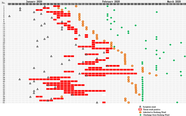
This retrospective study includes 51 patients with mild-to-moderate COVID-19 (median age: 34.0 years and 47.1% male) between January 31 and February 28, 2020. Demographic, clinical, laboratory, and computed tomography (CT) imaging data were collected before and after two consecutive negative respiratory SARS-CoV-2 tests.

RESULTS

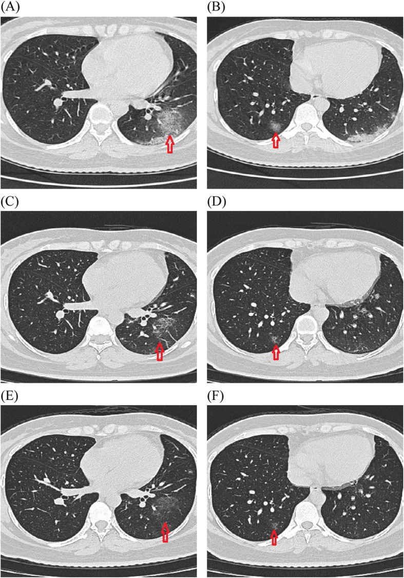
Following a negative test result, the patients' clinical symptoms continued to recover, but abnormal imaging findings were observed in all moderate cases. Specifically, 77.4% of patients with moderate COVID-19 exhibited multi-lobar lung involvement and lesions were more frequently observed in the lower lobes. The most common CT imaging manifestations were ground-glass opacities (51.6%) and fibrous stripes (54.8%%). Twelve of the 31 patients with moderate COVID-19 underwent repeated chest CT scans after a negative SARS-CoV-2 test. Among them, the ground-glass opacities decreased by > 60% within 1 week in seven patients (58.3%), but by < 5% in four patients (13.8%).

CONCLUSIONS

Following a positive and subsequent negative SARS-CoV-2 tests, patients with COVID-19 continued to recover despite exhibiting persistent clinical symptoms and abnormal imaging findings.

摘要

背景

大量研究已经探讨了严重急性呼吸综合征冠状病毒 2(SARS-CoV-2)感染患者进展为 2019 年冠状病毒病(COVID-19)的临床和影像学特征。然而,对于感染消退后呼吸道核酸检测结果为阴性的患者,这些特征知之甚少。在本研究中,我们旨在描述一组中国患者的这些特征。

方法

这项回顾性研究纳入了 2020 年 1 月 31 日至 2 月 28 日期间 51 例轻度至中度 COVID-19 患者(中位年龄:34.0 岁,47.1%为男性)。收集了两次连续呼吸道 SARS-CoV-2 检测均为阴性前后的人口统计学、临床、实验室和计算机断层扫描(CT)影像学数据。

结果

在检测结果为阴性后,患者的临床症状继续恢复,但所有中度病例均观察到异常影像学表现。具体而言,77.4%的中度 COVID-19 患者存在多肺叶受累,病变更常发生在下肺叶。最常见的 CT 影像学表现为磨玻璃影(51.6%)和纤维条纹(54.8%)。31 例中度 COVID-19 患者中有 12 例在 SARS-CoV-2 检测结果为阴性后接受了重复胸部 CT 扫描。其中,7 例(58.3%)患者的磨玻璃影在 1 周内减少了 >60%,但 4 例(13.8%)患者减少了 <5%。

结论

在 SARS-CoV-2 检测结果由阳性转为阴性后,COVID-19 患者尽管持续存在临床症状和异常影像学表现,但仍继续恢复。

https://cdn.ncbi.nlm.nih.gov/pmc/blobs/06dc/8034079/76463e0beac5/12879_2021_6013_Fig2_HTML.jpg
https://cdn.ncbi.nlm.nih.gov/pmc/blobs/06dc/8034079/6b1b70dbf8c5/12879_2021_6013_Fig1_HTML.jpg
https://cdn.ncbi.nlm.nih.gov/pmc/blobs/06dc/8034079/76463e0beac5/12879_2021_6013_Fig2_HTML.jpg
https://cdn.ncbi.nlm.nih.gov/pmc/blobs/06dc/8034079/6b1b70dbf8c5/12879_2021_6013_Fig1_HTML.jpg
https://cdn.ncbi.nlm.nih.gov/pmc/blobs/06dc/8034079/76463e0beac5/12879_2021_6013_Fig2_HTML.jpg

相似文献

1
Clinical and CT features of mild-to-moderate COVID-19 cases after two sequential negative nucleic acid testing results: a retrospective analysis.两次连续核酸检测阴性后轻症至普通型 COVID-19 病例的临床和 CT 特征:一项回顾性分析。
BMC Infect Dis. 2021 Apr 8;21(1):333. doi: 10.1186/s12879-021-06013-x.
2
Chest Computed Tomography and Clinical Follow-Up of Discharged Patients with COVID-19 in Wenzhou City, Zhejiang, China.中国浙江省温州市出院 COVID-19 患者的胸部计算机断层扫描和临床随访。
Ann Am Thorac Soc. 2020 Oct;17(10):1231-1237. doi: 10.1513/AnnalsATS.202004-324OC.
3
Temporal relationship between serial RT-PCR results and serial chest CT imaging, and serial CT changes in coronavirus 2019 (COVID-19) pneumonia: a descriptive study of 155 cases in China.新型冠状病毒(COVID-19)肺炎的实时逆转录聚合酶链反应(RT-PCR)结果与系列胸部 CT 影像学及系列 CT 改变的时间关系:中国 155 例的描述性研究。
Eur Radiol. 2021 Mar;31(3):1175-1184. doi: 10.1007/s00330-020-07268-9. Epub 2020 Sep 15.
4
Imaging and clinical features of patients with 2019 novel coronavirus SARS-CoV-2.新型冠状病毒 2019 年 SARS-CoV-2 患者的影像学和临床特征。
Eur J Nucl Med Mol Imaging. 2020 May;47(5):1275-1280. doi: 10.1007/s00259-020-04735-9. Epub 2020 Feb 28.
5
[Epidemiological and clinical features of children with mild coronavirus disease 2019].[2019冠状病毒病轻症儿童的流行病学和临床特征]
Zhongguo Dang Dai Er Ke Za Zhi. 2021 May;23(5):460-465. doi: 10.7499/j.issn.1008-8830.2101175.
6
CT-scan findings of COVID-19 pneumonia based on the time elapsed from the beginning of symptoms to the CT imaging evaluation: a descriptive study in Iran.基于从症状出现到 CT 影像评估时间的 COVID-19 肺炎 CT 扫描结果:伊朗的一项描述性研究。
Rom J Intern Med. 2020 Dec 17;58(4):242-250. doi: 10.2478/rjim-2020-0019. Print 2020 Dec 1.
7
[Clinical features and high resolution CT imaging evolution of coronavirus disease 2019].新型冠状病毒肺炎的临床特征及高分辨率CT影像学演变
Zhonghua Jie He He Hu Xi Za Zhi. 2020 Jun 12;43(6):509-515. doi: 10.3760/cma.j.cn112147-20200214-00094.
8
A comparison of clinical, laboratory and chest CT findings of laboratory-confirmed and clinically diagnosed COVID-19 patients at first admission.比较首次入院时经实验室确诊和临床诊断的 COVID-19 患者的临床、实验室和胸部 CT 表现。
Diagn Interv Radiol. 2021 May;27(3):336-343. doi: 10.5152/dir.2020.20270.
9
Clinical Features and Chest CT Manifestations of Coronavirus Disease 2019 (COVID-19) in a Single-Center Study in Shanghai, China.中国上海单中心研究的 2019 年冠状病毒病(COVID-19)的临床特征和胸部 CT 表现。
AJR Am J Roentgenol. 2020 Jul;215(1):121-126. doi: 10.2214/AJR.20.22959. Epub 2020 Mar 14.
10
[Analysis of CT features of 15 children with 2019 novel coronavirus infection].[15例2019新型冠状病毒感染儿童的CT特征分析]
Zhonghua Er Ke Za Zhi. 2020 Apr 2;58(4):275-278. doi: 10.3760/cma.j.cn112140-20200210-00071.

引用本文的文献

1
Chest Computed Tomography Is an Efficient Method for Initial Diagnosis of COVID-19: An Observational Study.胸部计算机断层扫描是COVID-19初始诊断的有效方法:一项观察性研究。
Front Med (Lausanne). 2022 Apr 12;9:848656. doi: 10.3389/fmed.2022.848656. eCollection 2022.
2
The efficacy and adverse effects of favipiravir on patients with COVID-19: A systematic review and meta-analysis of published clinical trials and observational studies.法匹拉韦治疗 COVID-19 患者的疗效和不良反应:基于已发表临床试验和观察性研究的系统评价和荟萃分析。
Int J Infect Dis. 2022 Jul;120:217-227. doi: 10.1016/j.ijid.2022.04.035. Epub 2022 Apr 22.
3

本文引用的文献

1
Chest CT in patients with a moderate or high pretest probability of COVID-19 and negative swab.胸部 CT 检查在中度或高度疑似 COVID-19 且咽拭子检测阴性的患者中的应用。
Radiol Med. 2020 Dec;125(12):1260-1270. doi: 10.1007/s11547-020-01269-w. Epub 2020 Aug 29.
2
CT features of COVID-19 patients with two consecutive negative RT-PCR tests after treatment.治疗后连续两次 RT-PCR 检测阴性的 COVID-19 患者的 CT 特征。
Sci Rep. 2020 Jul 14;10(1):11548. doi: 10.1038/s41598-020-68509-x.
3
Chest CT of COVID-19 in patients with a negative first RT-PCR test: Comparison with patients with a positive first RT-PCR test.
A rapid RNA extraction-free lateral flow assay for molecular point-of-care detection of SARS-CoV-2 augmented by chemical probes.
一种基于化学探针的免 RNA 提取侧向流检测方法,可快速进行 SARS-CoV-2 的分子即时检测。
Biosens Bioelectron. 2022 Mar 15;200:113900. doi: 10.1016/j.bios.2021.113900. Epub 2021 Dec 18.
首次逆转录聚合酶链反应(RT-PCR)检测结果为阴性的COVID-19患者的胸部CT:与首次RT-PCR检测结果为阳性的患者比较。
Medicine (Baltimore). 2020 Jun 26;99(26):e20837. doi: 10.1097/MD.0000000000020837.
4
Clinical characteristics of coronavirus disease 2019 (COVID-19) in China: A systematic review and meta-analysis.《中国 2019 年冠状病毒病(COVID-19)的临床特征:系统评价和荟萃分析》
J Infect. 2020 Jun;80(6):656-665. doi: 10.1016/j.jinf.2020.03.041. Epub 2020 Apr 10.
5
Clinical and computed tomographic (CT) images characteristics in the patients with COVID-19 infection: What should radiologists need to know?新型冠状病毒肺炎(COVID-19)感染患者的临床和计算机断层扫描(CT)图像特征:放射科医生需要了解什么?
J Xray Sci Technol. 2020;28(3):369-381. doi: 10.3233/XST-200670.
6
Imaging and clinical features of patients with 2019 novel coronavirus SARS-CoV-2: A systematic review and meta-analysis.新型冠状病毒 SARS-CoV-2 感染患者的影像学和临床特征:系统评价和荟萃分析。
J Med Virol. 2020 Sep;92(9):1449-1459. doi: 10.1002/jmv.25822. Epub 2020 Apr 10.
7
Novel Coronavirus Infection (COVID-19) in Humans: A Scoping Review and Meta-Analysis.人类新型冠状病毒感染(COVID-19):一项范围综述与荟萃分析
J Clin Med. 2020 Mar 30;9(4):941. doi: 10.3390/jcm9040941.
8
Quantitative Detection and Viral Load Analysis of SARS-CoV-2 in Infected Patients.新型冠状病毒感染患者的病毒定量检测与载量分析
Clin Infect Dis. 2020 Jul 28;71(15):793-798. doi: 10.1093/cid/ciaa345.
9
Prolonged presence of SARS-CoV-2 viral RNA in faecal samples.严重急性呼吸综合征冠状病毒2(SARS-CoV-2)病毒RNA在粪便样本中的长期存在。
Lancet Gastroenterol Hepatol. 2020 May;5(5):434-435. doi: 10.1016/S2468-1253(20)30083-2. Epub 2020 Mar 20.
10
Clinical, laboratory and imaging features of COVID-19: A systematic review and meta-analysis.新型冠状病毒肺炎的临床、实验室和影像学特征:系统评价和荟萃分析。
Travel Med Infect Dis. 2020 Mar-Apr;34:101623. doi: 10.1016/j.tmaid.2020.101623. Epub 2020 Mar 13.