Image Data Sciences, Definiens GmbH, Munich, Germany.
Department of Pathology, GROW School for Oncology and Developmental Biology, Maastricht University Medical Center+, Maastricht, The Netherlands.
J Pathol Clin Res. 2020 Oct;6(4):273-282. doi: 10.1002/cjp2.170. Epub 2020 Jun 27.
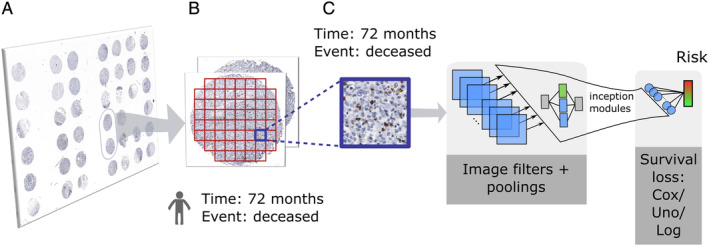
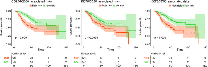
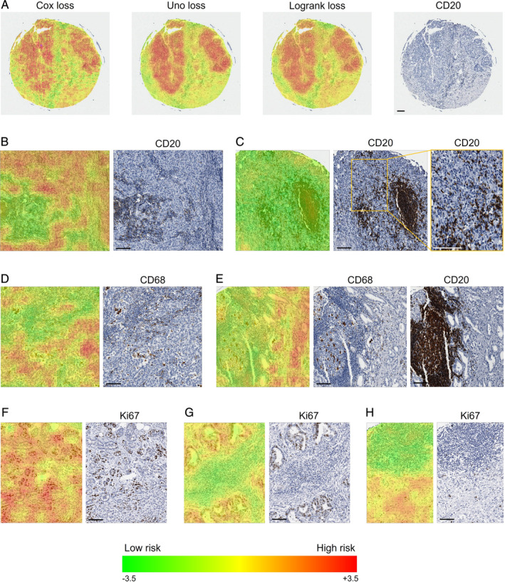
The biological complexity reflected in histology images requires advanced approaches for unbiased prognostication. Machine learning and particularly deep learning methods are increasingly applied in the field of digital pathology. In this study, we propose new ways to predict risk for cancer-specific death from digital images of immunohistochemically (IHC) stained tissue microarrays (TMAs). Specifically, we evaluated a cohort of 248 gastric cancer patients using convolutional neural networks (CNNs) in an end-to-end weakly supervised scheme independent of subjective pathologist input. To account for the time-to-event characteristic of the outcome data, we developed new survival models to guide the network training. In addition to the standard H&E staining, we investigated the prognostic value of a panel of immune cell markers (CD8, CD20, CD68) and a proliferation marker (Ki67). Our CNN-derived risk scores provided additional prognostic value when compared to the gold standard prognostic tool TNM stage. The CNN-derived risk scores were also shown to be superior when systematically compared to cell density measurements or a CNN score derived from binary 5-year survival classification, which ignores time-to-event. To better understand the underlying biological mechanisms, we qualitatively investigated risk heat maps for each marker which visualised the network output. We identified patterns of biological interest that were related to low risk of cancer-specific death such as the presence of B-cell predominated clusters and Ki67 positive sub-regions and showed that the corresponding risk scores had prognostic value in multivariate Cox regression analyses (Ki67&CD20 risks: hazard ratio (HR) = 1.47, 95% confidence interval (CI) = 1.15-1.89, p = 0.002; CD20&CD68 risks: HR = 1.33, 95% CI = 1.07-1.67, p = 0.009). Our study demonstrates the potential additional value that deep learning in combination with a panel of IHC markers can bring to the field of precision oncology.
组织学图像中反映的生物学复杂性需要采用先进的方法进行无偏预后预测。机器学习,特别是深度学习方法,在数字病理学领域得到了越来越多的应用。在这项研究中,我们提出了从免疫组织化学(IHC)染色组织微阵列(TMA)的数字图像预测癌症特异性死亡风险的新方法。具体来说,我们使用卷积神经网络(CNN)在无需主观病理学家输入的端到端弱监督方案中评估了 248 例胃癌患者队列。为了说明结果数据的时变特征,我们开发了新的生存模型来指导网络训练。除了标准的 H&E 染色外,我们还研究了一组免疫细胞标志物(CD8、CD20、CD68)和增殖标志物(Ki67)的预后价值。与金标准预后工具 TNM 分期相比,我们的 CNN 衍生风险评分提供了额外的预后价值。当系统地与细胞密度测量或源自二元 5 年生存分类的 CNN 评分进行比较时,CNN 衍生风险评分也显示出优越性,而后者忽略了时变。为了更好地了解潜在的生物学机制,我们对每个标志物的风险热图进行了定性研究,这些热图可视化了网络输出。我们确定了与癌症特异性死亡风险低相关的感兴趣的生物学模式,例如 B 细胞为主的簇和 Ki67 阳性亚区的存在,并表明相应的风险评分在多变量 Cox 回归分析中具有预后价值(Ki67&CD20 风险:风险比(HR)= 1.47,95%置信区间(CI)= 1.15-1.89,p = 0.002;CD20&CD68 风险:HR = 1.33,95% CI = 1.07-1.67,p = 0.009)。我们的研究表明,深度学习与一组 IHC 标志物相结合可以为精准肿瘤学领域带来潜在的附加价值。