Tsai Chih-Mong, Wu Fang-Yu, Chai Jyh-Wen, Chen Mu-Hsiung, Kao Chih-Ting
Department of Dentistry, National Taiwan University Hospital, Taipei, Taiwan.
Department of Radiology, Taichung Veterans General Hospital, Taichung, Taiwan.
J Dent Sci. 2020 Jun;15(2):153-162. doi: 10.1016/j.jds.2020.03.004. Epub 2020 Apr 10.
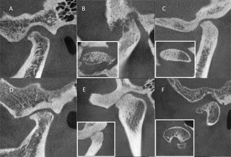
BACKGROUND/PURPOSE: The clinical diagnosis of temporomandibular joint (TMJ) degenerative joint disease (DJD) is based primarily on radiographic features of the condyle and articular eminence. The purpose of this study was to compare the reliability, sensitivity, and specificity of using plain radiography to that of cone-beam computerized tomography (CBCT) in identifying different types of osseous degenerative features in the TMJ condyle.
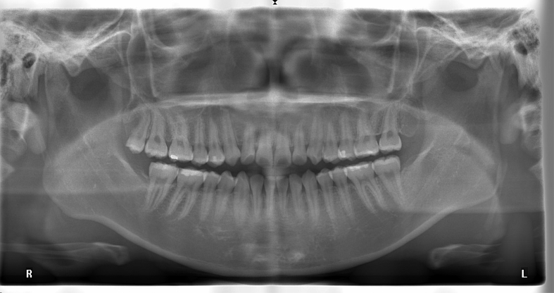
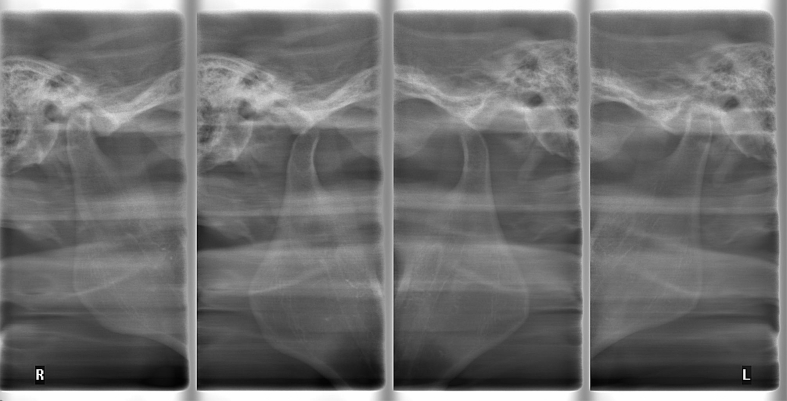
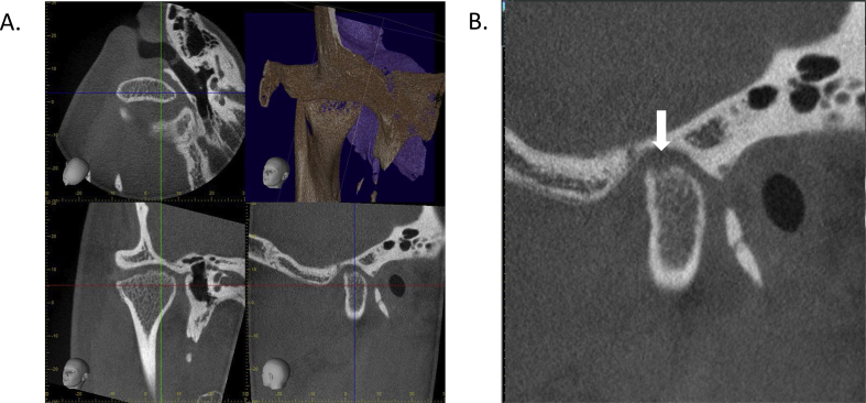
The panoramic radiography (PANO), TMJ quadruple radiography (TMJQR) and CBCT images of 29 patients' TMJs were retrieved from a computer database and independently evaluated by a young oral surgeon and a senior TMD specialist. The examiners diagnosed osseous degenerative features on the radiographic images. The radiologist-assisted CBCT diagnoses were used as a reference standard and the reliability, sensitivity, and specificity of using the three radiographic modalities were statistically analyzed.
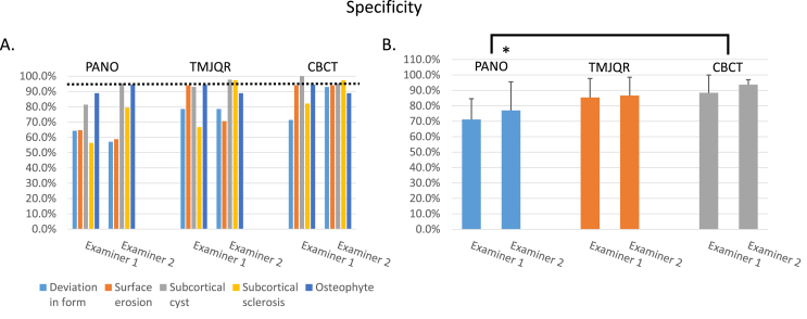
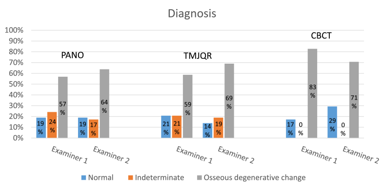
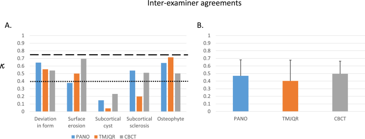
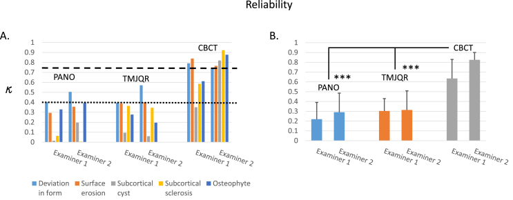
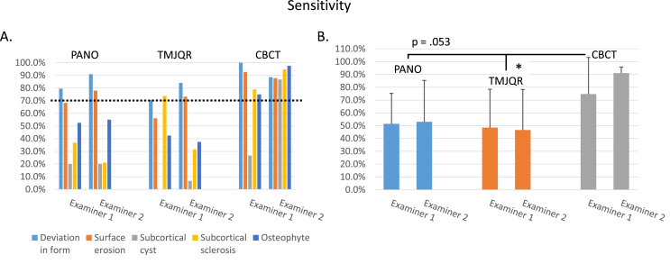
There were cases of indeterminate diagnoses using the PANO and TMJQR due to superimposition from surrounding structures, but none using CBCT. Reliability was generally poor when using PANO and TMJQR for detecting osseous degenerative features of the TMJ condyle but good to excellent when using CBCT. The sensitivity and specificity in the use of PANO and TMJQR were typically below acceptable, but the levels were generally satisfactory when using CBCT.
CBCT is superior to plain radiographic modalities for diagnosing osseous degenerative features of TMJs with regard to indeterminate cases, reliability, sensitivity, and specificity. It is recommended that CBCT can be used as an effective tool in identifying TMJ osteoarthritis.
背景/目的:颞下颌关节(TMJ)退行性关节病(DJD)的临床诊断主要基于髁突和关节结节的影像学特征。本研究的目的是比较使用普通X线摄影与锥形束计算机断层扫描(CBCT)在识别TMJ髁突不同类型骨退行性特征方面的可靠性、敏感性和特异性。
从计算机数据库中检索29例患者TMJ的全景X线片(PANO)、TMJ四联X线片(TMJQR)和CBCT图像,并由一名年轻口腔外科医生和一名资深颞下颌关节紊乱病(TMD)专家独立评估。检查者在影像学图像上诊断骨退行性特征。以放射科医生辅助的CBCT诊断作为参考标准,并对三种影像学检查方法的可靠性、敏感性和特异性进行统计学分析。
由于周围结构的重叠,使用PANO和TMJQR时有不确定诊断的病例,但使用CBCT时没有。使用PANO和TMJQR检测TMJ髁突的骨退行性特征时,可靠性一般较差,但使用CBCT时可靠性良好至优秀。使用PANO和TMJQR时的敏感性和特异性通常低于可接受水平,但使用CBCT时一般令人满意。
在不确定病例、可靠性、敏感性和特异性方面,CBCT在诊断TMJ的骨退行性特征方面优于普通X线检查方法。建议将CBCT用作识别TMJ骨关节炎的有效工具。