Spine Center, Department of Orthopaedics, Shanghai Changzheng Hospital, Second Military Medical University, Shanghai, 200003, China.
Department of Endocrinology and Metabolism, Shanghai Diabetes Institute, Shanghai Key Laboratory of Diabetes Mellitus, Shanghai Clinical Center for Diabetes, Shanghai Jiao Tong University Affiliated Sixth People's Hospital, Shanghai, 200233, China.
Stem Cell Res Ther. 2020 Jun 29;11(1):259. doi: 10.1186/s13287-020-01756-x.
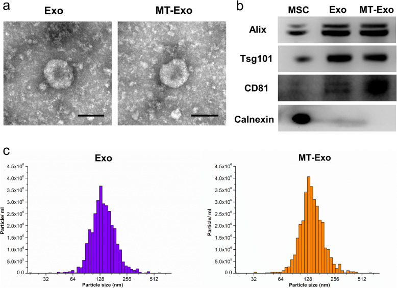
After surgery, wound recovery in diabetic patients may be disrupted due to delayed inflammation, which can lead to undesired consequences, and there is currently a lack of effective measures to address this issue. Mesenchymal stem cell (MSC)-derived exosomes (Exo) have been proven to be appropriate candidates for diabetic wound healing through the anti-inflammatory effects. In this study, we investigated whether melatonin (MT)-pretreated MSCs-derived exosomes (MT-Exo) could exert superior effects on diabetic wound healing, and we attempted to elucidate the underlying mechanism.
For the evaluation of the anti-inflammatory effect of MT-Exo, in vitro and in vivo studies were performed. For in vitro research, we detected the secreted levels of inflammation-related factors, such as IL-1β, TNF-α and IL-10 via ELISA and the relative gene expression of the IL-1β, TNF-α, IL-10, Arg-1 and iNOS via qRT-PCR and investigated the expression of PTEN, AKT and p-AKT by Western blotting. For in vivo study, we established air pouch model and streptozotocin (STZ)-treated diabetic wound model, and evaluated the effect of MT-Exo by flow cytometry, optical imaging, H&E staining, Masson trichrome staining, immunohistochemical staining, immunofluorescence, and qRT-PCR (α-SMA, collagen I and III).
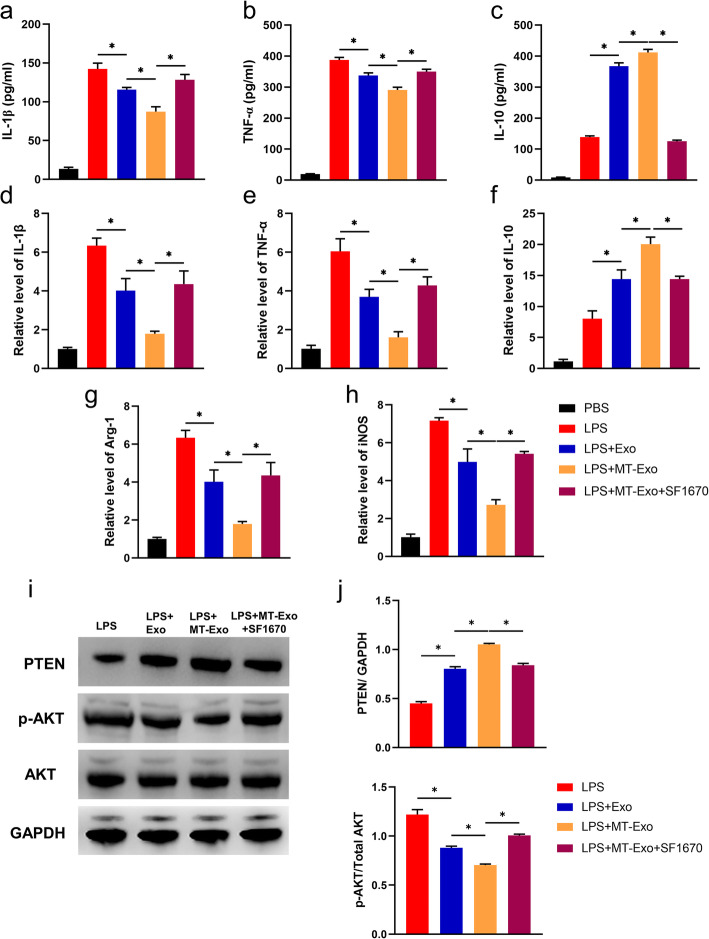
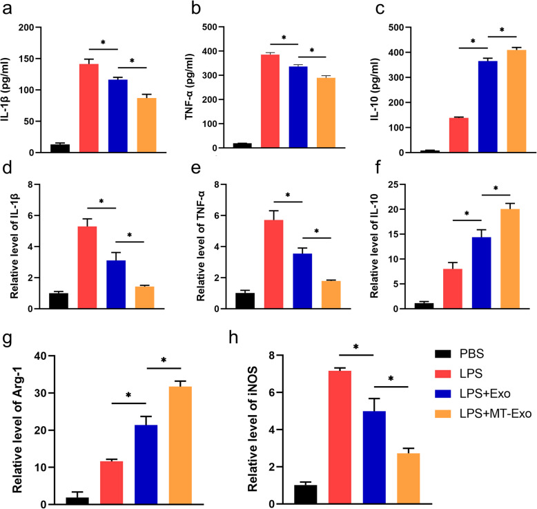
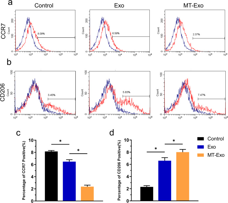
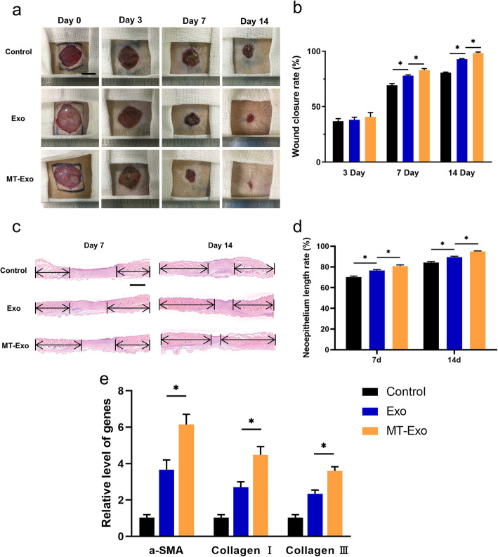
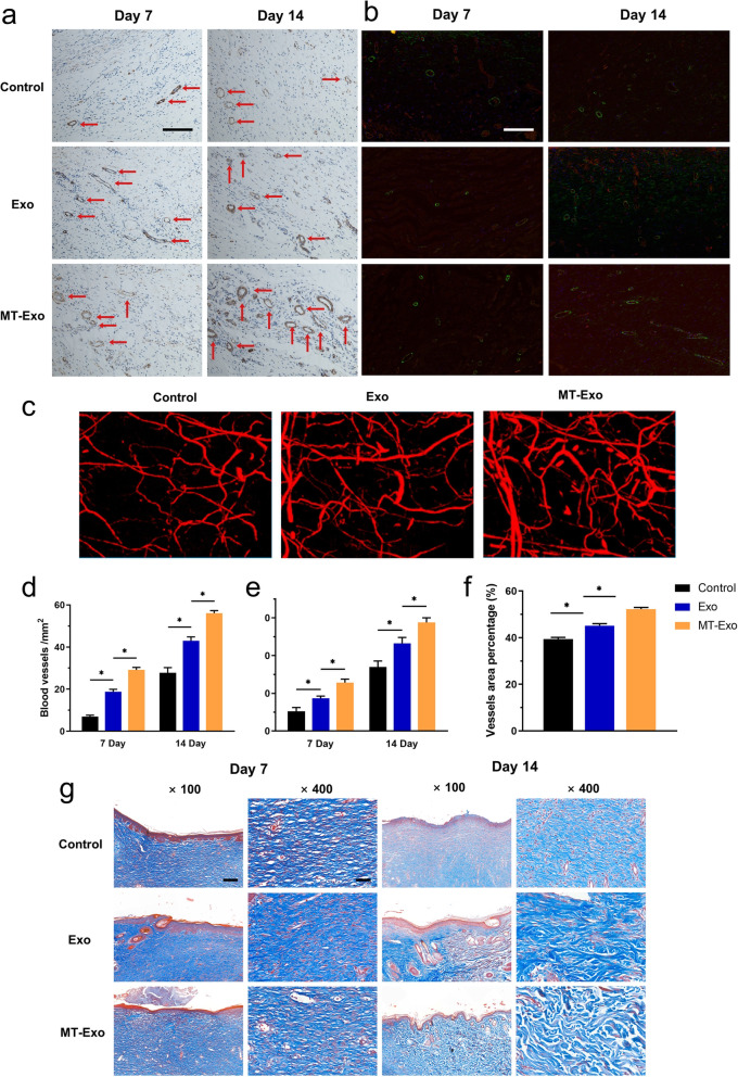
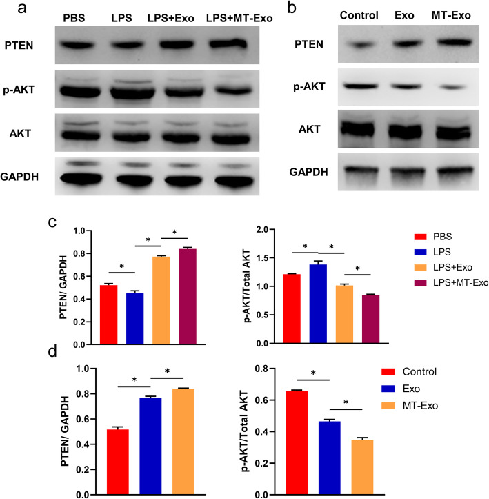
MT-Exo significantly suppressed the pro-inflammatory factors IL-1β and TNF-α and reduced the relative gene expression of IL-1β, TNF-α and iNOS, while promoting the anti-inflammatory factor IL-10 along with increasing the relative expression of IL-10 and Arg-1, compared with that of the PBS, LPS and the Exo groups in vitro. This effect was mediated by the increased ratio of M2 polarization to M1 polarization through upregulating the expression of PTEN and inhibiting the phosphorylation of AKT. Similarly, MT-Exo significantly promoted the healing of diabetic wounds by inhibiting inflammation, thereby further facilitating angiogenesis and collagen synthesis in vivo.
MT-Exo could promote diabetic wound healing by suppressing the inflammatory response, which was achieved by increasing the ratio of M2 polarization to M1 polarization through activating the PTEN/AKT signalling pathway, and the pretreatment of MT was proved to be a promising method for treating diabetic wound healing.
糖尿病患者手术后,由于炎症反应延迟,伤口恢复可能会受到干扰,从而导致不理想的后果,而目前缺乏有效的治疗措施。间充质干细胞(MSC)衍生的外泌体(Exo)已被证明通过抗炎作用是治疗糖尿病伤口愈合的合适候选物。在这项研究中,我们研究了褪黑素(MT)预处理的 MSC 衍生外泌体(MT-Exo)是否对糖尿病伤口愈合有更好的效果,并试图阐明其潜在机制。
为了评估 MT-Exo 的抗炎作用,进行了体外和体内研究。体外研究中,我们通过 ELISA 检测炎症相关因子如 IL-1β、TNF-α 和 IL-10 的分泌水平,通过 qRT-PCR 检测 IL-1β、TNF-α、IL-10、Arg-1 和 iNOS 的相对基因表达,通过 Western blot 检测 PTEN、AKT 和 p-AKT 的表达。在体内研究中,我们建立了气囊模型和链脲佐菌素(STZ)诱导的糖尿病伤口模型,通过流式细胞术、光学成像、H&E 染色、Masson 三色染色、免疫组织化学染色、免疫荧光和 qRT-PCR(α-SMA、胶原蛋白 I 和 III)评估 MT-Exo 的作用。
MT-Exo 显著抑制了促炎因子 IL-1β 和 TNF-α 的表达,并降低了 IL-1β、TNF-α 和 iNOS 的相对基因表达,同时促进了抗炎因子 IL-10 的表达,并增加了 IL-10 和 Arg-1 的相对表达,与 PBS、LPS 和 Exo 组相比。这种作用是通过上调 PTEN 的表达和抑制 AKT 的磷酸化来增加 M2 极化与 M1 极化的比例来介导的。同样,MT-Exo 还通过抑制炎症显著促进了糖尿病伤口的愈合,从而进一步促进了体内的血管生成和胶原合成。
MT-Exo 通过抑制炎症反应促进糖尿病伤口愈合,这是通过激活 PTEN/AKT 信号通路增加 M2 极化与 M1 极化的比例来实现的,而 MT 的预处理被证明是治疗糖尿病伤口愈合的一种有前途的方法。