Ahn Byeong-Seob, Oh Song Hee, Heo Chong-Kwan, Kim Gyu-Tae, Choi Yong-Suk, Hwang Eui-Hwan
Department of Oral and Maxillofacial Radiology, School of Dentistry, Kyung Hee University, Seoul, Korea.
School of Dentistry, Kyung Hee University, Seoul, Korea.
Imaging Sci Dent. 2020 Jun;50(2):125-132. doi: 10.5624/isd.2020.50.2.125. Epub 2020 Jun 18.
The positions of the mandibular foramen (MnF) and the lingula affect the success rate of inferior alveolar nerve block. The objective of this study was to investigate aspects of the MnF and the lingula relevant for mandibular block anesthesia using cone-beam computed tomography (CBCT).
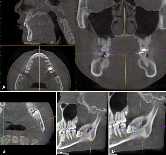
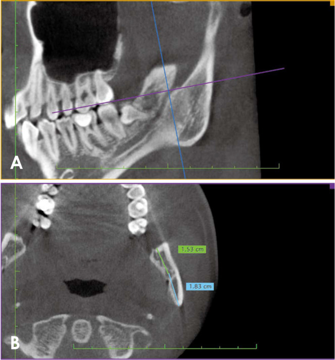
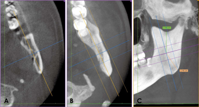
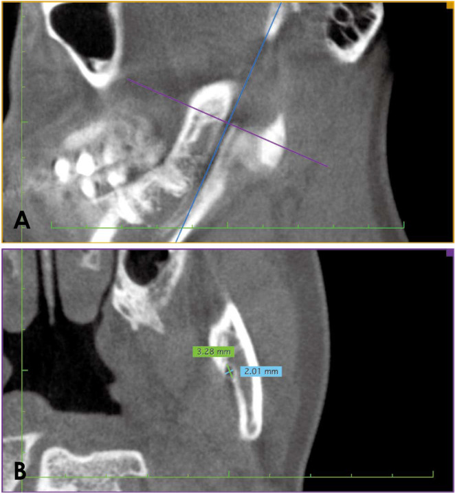
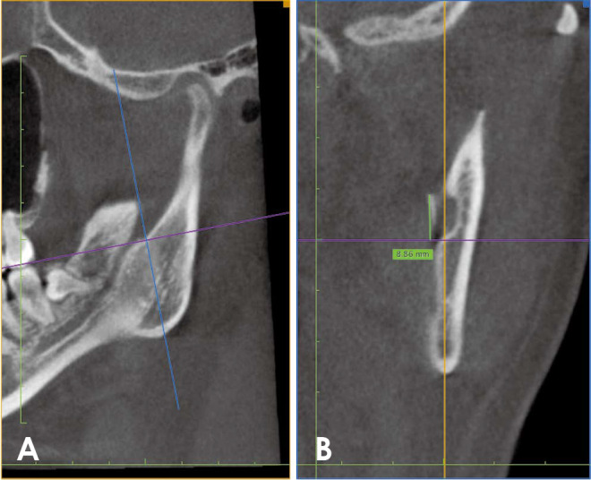
Fifty CBCT scans were collected from a picture archiving and communications system. All scans were taken using an Alphard Vega 3030 (Asahi Roentgen Co. Ltd., Kyoto, Japan). Fifty-eight MnFs of 30 subjects were included in the study. The position of the MnF, the size of the MnF, the position of the lingula, the size of the lingula, and the shape of the lingula were measured and recorded. All data were statistically analyzed at a significance level of <0.05.
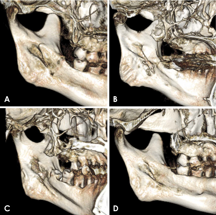
The position of MnF was 0.1 mm and 0.8 mm below the occlusal plane in males and females, respectively. The horizontal position of the MnF was slightly anterior to the center of the ramus in males and in the center in females (<0.05). The vertical position of the MnF was lower in females than in males (<0.05). The MnF was an oval shape with a longer anteroposterior dimension. The height of the lingula was 9.3 mm in males and 8.2 mm in females. The nodular type was the most common shape of the lingula, followed by the triangular, truncated, and assimilated types.
CBCT provided useful information about the MnF and lingula. This information could improve the success rate of mandibular blocks.
下颌孔(MnF)和舌骨的位置会影响下牙槽神经阻滞的成功率。本研究的目的是使用锥形束计算机断层扫描(CBCT)研究与下颌阻滞麻醉相关的下颌孔和舌骨的各个方面。
从图像存档与通信系统收集了50份CBCT扫描图像。所有扫描均使用Alphard Vega 3030(日本京都旭光电子有限公司)进行。30名受试者的58个下颌孔纳入研究。测量并记录下颌孔的位置、下颌孔的大小、舌骨的位置、舌骨的大小以及舌骨的形状。所有数据均在显著性水平<0.05下进行统计分析。
男性和女性下颌孔的位置分别在咬合平面以下0.1毫米和0.8毫米处。男性下颌孔的水平位置略靠前于下颌支中心,女性则位于中心(<0.05)。女性下颌孔的垂直位置低于男性(<0.05)。下颌孔呈椭圆形,前后径较长。男性舌骨的高度为9.3毫米,女性为8.2毫米。结节型是舌骨最常见的形状,其次是三角形、截断型和同化型。
CBCT提供了有关下颌孔和舌骨的有用信息。这些信息可以提高下颌阻滞的成功率。