National Medical Research Center for Obstetrics, Gynecology and Perinatology named after Academician V.I. Kulakov of the Ministry of Healthcare of Russian Federation, 117997 Moscow, Russia.
Moscow Institute of Physics and Technology, 141701 Moscow, Russia.
Int J Mol Sci. 2020 Jun 26;21(12):4568. doi: 10.3390/ijms21124568.
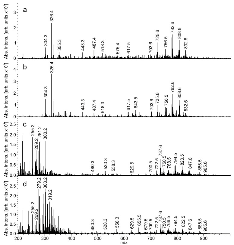
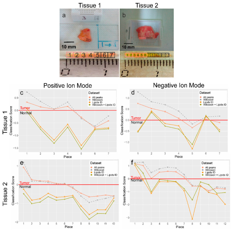
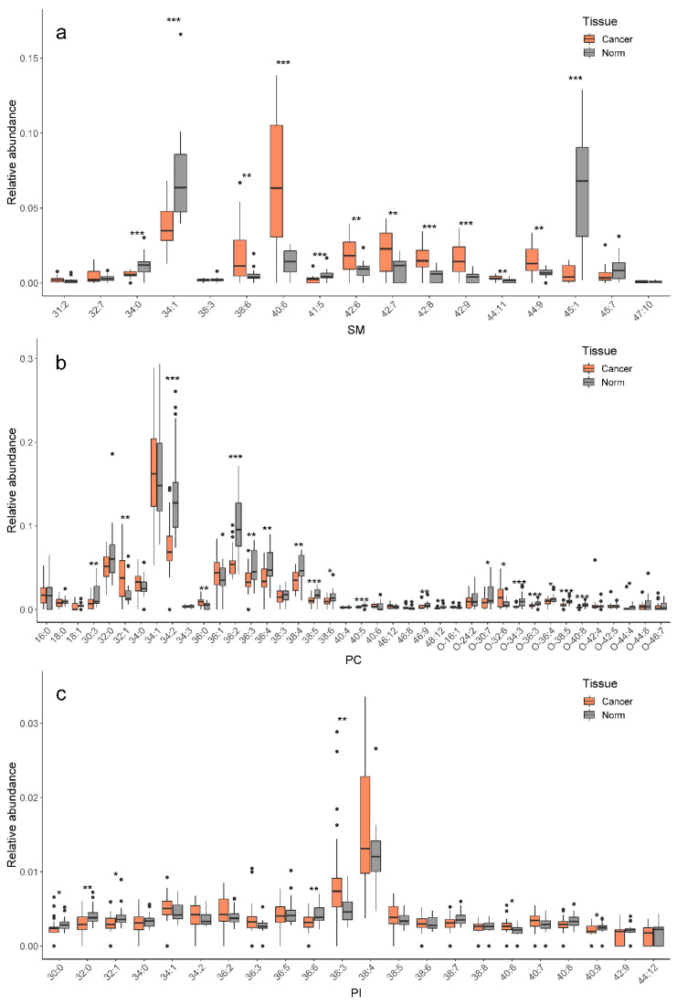
Current methods for the intraoperative determination of breast cancer margins commonly suffer from the insufficient accuracy, specificity and/or low speed of analysis, increasing the time and cost of operation as well the risk of cancer recurrence. The purpose of this study is to develop a method for the rapid and accurate determination of breast cancer margins using direct molecular profiling by mass spectrometry (MS). Direct molecular fingerprinting of tiny pieces of breast tissue (approximately 1 × 1 × 1 mm) is performed using a home-built tissue spray ionization source installed on a Maxis Impact quadrupole time-of-flight mass spectrometer (qTOF MS) (Bruker Daltonics, Hamburg, Germany). Statistical analysis of MS data from 50 samples of both normal and cancer tissue (from 25 patients) was performed using orthogonal projections onto latent structures discriminant analysis (OPLS-DA). Additionally, the results of OPLS classification of new 19 pieces of two tissue samples were compared with the results of histological analysis performed on the same tissues samples. The average time of analysis for one sample was about 5 min. Positive and negative ionization modes are used to provide complementary information and to find out the most informative method for a breast tissue classification. The analysis provides information on 11 lipid classes. OPLS-DA models are created for the classification of normal and cancer tissue based on the various datasets: All mass spectrometric peaks over 300 counts; peaks with a statistically significant difference of intensity determined by the Mann-Whitney U-test ( < 0.05); peaks identified as lipids; both identified and significantly different peaks. The highest values of Q2 have models built on all MS peaks and on significantly different peaks. While such models are useful for classification itself, they are of less value for building explanatory mechanisms of pathophysiology and providing a pathway analysis. Models based on identified peaks are preferable from this point of view. Results obtained by OPLS-DA classification of the tissue spray MS data of a new sample set ( = 19) revealed 100% sensitivity and specificity when compared to histological analysis, the "gold" standard for tissue classification. "All peaks" and "significantly different peaks" datasets in the positive ion mode were ideal for breast cancer tissue classification. Our results indicate the potential of tissue spray mass spectrometry for rapid, accurate and intraoperative diagnostics of breast cancer tissue as a means to reduce surgical intervention.
目前,用于术中确定乳腺癌边界的方法通常存在准确性、特异性和/或分析速度不足的问题,这增加了手术的时间和成本,以及癌症复发的风险。本研究旨在开发一种使用质谱(MS)直接分子谱快速准确地确定乳腺癌边界的方法。使用安装在 Maxis Impact 四极杆飞行时间质谱仪(qTOF MS)(Bruker Daltonics,德国汉堡)上的自制组织喷雾电离源对小块乳腺组织(约 1×1×1 毫米)进行直接分子指纹图谱分析。对来自 25 名患者的 50 份正常和癌症组织样本的 MS 数据进行正交投影到潜在结构判别分析(OPLS-DA)的统计分析。此外,还将新的 19 个组织样本的 OPLS 分类结果与对相同组织样本进行的组织学分析结果进行了比较。一个样本的平均分析时间约为 5 分钟。正、负离子模式用于提供互补信息,并找到最适合乳腺组织分类的信息。该分析提供了 11 种脂质类别的信息。根据不同的数据集创建 OPLS-DA 模型,用于正常组织和癌症组织的分类:超过 300 个计数的所有质谱峰;通过曼-惠特尼 U 检验( < 0.05)确定强度有统计学差异的峰;鉴定为脂质的峰;已鉴定且差异显著的峰。基于所有 MS 峰和显著差异峰构建的模型具有最高的 Q2 值。虽然这些模型对于分类本身很有用,但对于构建病理生理学的解释机制和提供途径分析的价值较小。从这个角度来看,基于鉴定峰的模型是首选。当与组织学分析(组织分类的“金标准”)相比时,新样本集(n=19)的组织喷雾 MS 数据的 OPLS-DA 分类获得的结果具有 100%的灵敏度和特异性。正离子模式下的“所有峰”和“差异显著峰”数据集是乳腺癌组织分类的理想选择。我们的研究结果表明,组织喷雾质谱法具有快速、准确和术中诊断乳腺癌组织的潜力,可作为减少手术干预的手段。