• 文献检索
  • 文档翻译
  • 深度研究
  • 学术资讯
  • Suppr Zotero 插件Zotero 插件
  • 邀请有礼
  • 套餐&价格
  • 历史记录
应用&插件
Suppr Zotero 插件Zotero 插件浏览器插件Mac 客户端Windows 客户端微信小程序
定价
高级版会员购买积分包购买API积分包
服务
文献检索文档翻译深度研究API 文档MCP 服务
关于我们
关于 Suppr公司介绍联系我们用户协议隐私条款
关注我们

Suppr 超能文献

核心技术专利:CN118964589B侵权必究
粤ICP备2023148730 号-1Suppr @ 2026

文献检索

告别复杂PubMed语法,用中文像聊天一样搜索,搜遍4000万医学文献。AI智能推荐,让科研检索更轻松。

立即免费搜索

文件翻译

保留排版,准确专业,支持PDF/Word/PPT等文件格式,支持 12+语言互译。

免费翻译文档

深度研究

AI帮你快速写综述,25分钟生成高质量综述,智能提取关键信息,辅助科研写作。

立即免费体验

清醒大鼠静息态 fMRI 的开放数据库。

An open database of resting-state fMRI in awake rats.

机构信息

Department of Biomedical Engineering, The Pennsylvania State University, University Park, PA, 16802, USA.

Neuroscience Program, The Huck Institutes of the Life Sciences, The Pennsylvania State University, University Park, PA, 16802, USA.

出版信息

Neuroimage. 2020 Oct 15;220:117094. doi: 10.1016/j.neuroimage.2020.117094. Epub 2020 Jun 28.

DOI:10.1016/j.neuroimage.2020.117094
PMID:32610063
原文链接:https://pmc.ncbi.nlm.nih.gov/articles/PMC7605641/
Abstract

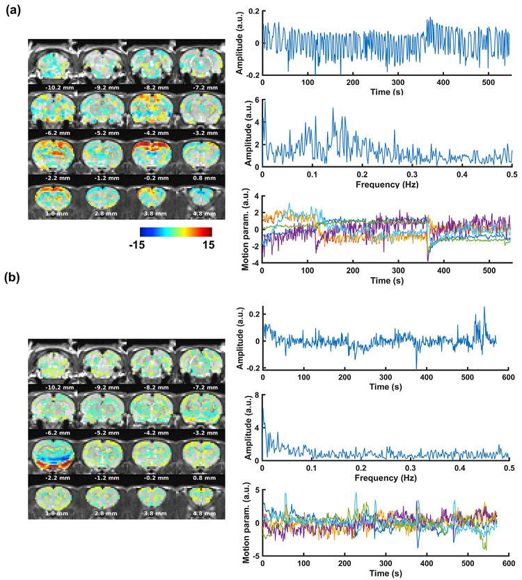
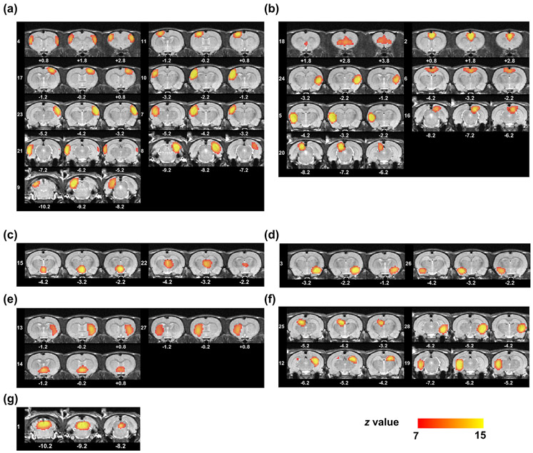
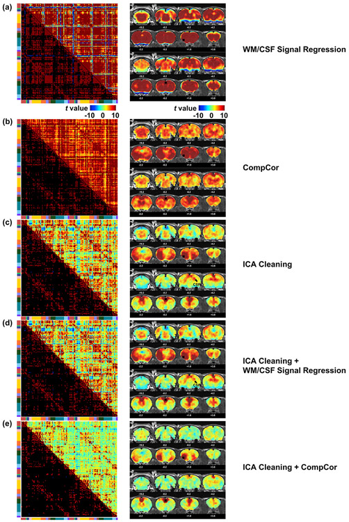
Rodent models are essential to translational research in health and disease. Investigation in rodent brain function and organization at the systems level using resting-state functional magnetic resonance imaging (rsfMRI) has become increasingly popular. Due to this rapid progress, publicly shared rodent rsfMRI databases can be of particular interest and importance to the scientific community, as inspired by human neuroscience and psychiatric research that are substantially facilitated by open human neuroimaging datasets. However, such databases in rats are still rare. In this paper, we share an open rsfMRI database acquired in 90 rats with a well-established awake imaging paradigm that avoids anesthesia interference. Both raw and preprocessed data are made publicly available. Procedures in data preprocessing to remove artefacts induced by the scanner, head motion and non-neural physiological noise are described in details. We also showcase inter-regional functional connectivity and functional networks obtained from the database.

摘要

啮齿动物模型对于健康和疾病的转化研究至关重要。使用静息态功能磁共振成像(rsfMRI)研究啮齿动物大脑功能和系统水平的组织已变得越来越流行。由于这一快速发展,受人类神经科学和精神病学研究的启发,公开共享的啮齿动物 rsfMRI 数据库可能对科学界特别感兴趣和重要,这些研究得益于开放的人类神经影像学数据集。然而,在大鼠中,这样的数据库仍然很少。在本文中,我们分享了一个使用经过充分验证的清醒成像范式获得的公开 rsfMRI 数据库,该范式避免了麻醉干扰。原始数据和预处理数据都可供公开使用。详细描述了数据预处理过程中去除由扫描仪、头部运动和非神经生理噪声引起的伪影的步骤。我们还展示了从数据库中获得的区域间功能连接和功能网络。

https://cdn.ncbi.nlm.nih.gov/pmc/blobs/f9d2/7605641/442013c6908e/nihms-1639062-f0009.jpg
https://cdn.ncbi.nlm.nih.gov/pmc/blobs/f9d2/7605641/ecee5da2c9e8/nihms-1639062-f0001.jpg
https://cdn.ncbi.nlm.nih.gov/pmc/blobs/f9d2/7605641/d514284f1ccd/nihms-1639062-f0002.jpg
https://cdn.ncbi.nlm.nih.gov/pmc/blobs/f9d2/7605641/adf07a671d4c/nihms-1639062-f0003.jpg
https://cdn.ncbi.nlm.nih.gov/pmc/blobs/f9d2/7605641/3876534af7c9/nihms-1639062-f0004.jpg
https://cdn.ncbi.nlm.nih.gov/pmc/blobs/f9d2/7605641/b5ef286dcdb5/nihms-1639062-f0005.jpg
https://cdn.ncbi.nlm.nih.gov/pmc/blobs/f9d2/7605641/43515ee10016/nihms-1639062-f0006.jpg
https://cdn.ncbi.nlm.nih.gov/pmc/blobs/f9d2/7605641/786b8adc1dee/nihms-1639062-f0007.jpg
https://cdn.ncbi.nlm.nih.gov/pmc/blobs/f9d2/7605641/61677734df29/nihms-1639062-f0008.jpg
https://cdn.ncbi.nlm.nih.gov/pmc/blobs/f9d2/7605641/442013c6908e/nihms-1639062-f0009.jpg

相似文献

1
An open database of resting-state fMRI in awake rats.清醒大鼠静息态 fMRI 的开放数据库。
Neuroimage. 2020 Oct 15;220:117094. doi: 10.1016/j.neuroimage.2020.117094. Epub 2020 Jun 28.
2
Gaining insight into the neural basis of resting-state fMRI signal.深入了解静息态 fMRI 信号的神经基础。
Neuroimage. 2022 Apr 15;250:118960. doi: 10.1016/j.neuroimage.2022.118960. Epub 2022 Feb 1.
3
Resting-state functional MRI of the default mode network in epilepsy.癫痫默认模式网络的静息态功能磁共振成像。
Epilepsy Behav. 2020 Oct;111:107308. doi: 10.1016/j.yebeh.2020.107308. Epub 2020 Jul 19.
4
An isotropic EPI database and analytical pipelines for rat brain resting-state fMRI.大鼠脑静息态 fMRI 的各向同性 EPI 数据库和分析管道。
Neuroimage. 2021 Nov;243:118541. doi: 10.1016/j.neuroimage.2021.118541. Epub 2021 Aug 31.
5
Resting-state functional magnetic resonance imaging versus task-based activity for language mapping and correlation with perioperative cortical mapping.静息态功能磁共振成像与任务态活动在语言定位中的比较,以及与围手术期皮质定位的相关性。
Brain Behav. 2019 Oct;9(10):e01362. doi: 10.1002/brb3.1362. Epub 2019 Sep 30.
6
Default mode network, motor network, dorsal and ventral basal ganglia networks in the rat brain: comparison to human networks using resting state-fMRI.大鼠脑中的默认模式网络、运动网络、背侧和腹侧基底神经节网络:与使用静息态功能磁共振成像的人类网络的比较。
PLoS One. 2015 Mar 19;10(3):e0120345. doi: 10.1371/journal.pone.0120345. eCollection 2015.
7
Biophysical and neural basis of resting state functional connectivity: Evidence from non-human primates.静息态功能连接的生物物理和神经基础:来自非人类灵长类动物的证据。
Magn Reson Imaging. 2017 Jun;39:71-81. doi: 10.1016/j.mri.2017.01.020. Epub 2017 Feb 2.
8
Global brain signal in awake rats.清醒大鼠的全球脑信号。
Brain Struct Funct. 2020 Jan;225(1):227-240. doi: 10.1007/s00429-019-01996-5. Epub 2019 Dec 4.
9
Biomarkers, designs, and interpretations of resting-state fMRI in translational pharmacological research: A review of state-of-the-Art, challenges, and opportunities for studying brain chemistry.转化药理学研究中静息态功能磁共振成像的生物标志物、设计与解读:脑化学研究的现状、挑战与机遇综述
Hum Brain Mapp. 2017 Apr;38(4):2276-2325. doi: 10.1002/hbm.23516. Epub 2017 Feb 1.
10
Abnormal functional connectivity and effective connectivity between the default mode network and attention networks in patients with alcohol-use disorder.酒精使用障碍患者的默认模式网络和注意网络之间的异常功能连接和有效连接。
Acta Radiol. 2021 Feb;62(2):251-259. doi: 10.1177/0284185120923270. Epub 2020 May 18.

引用本文的文献

1
Adolescent alcohol consumption alters sex-specific behaviors associated with prefrontal functional connectivity in mice.青少年饮酒会改变与小鼠前额叶功能连接相关的性别特异性行为。
bioRxiv. 2025 Jun 8:2025.06.05.658112. doi: 10.1101/2025.06.05.658112.
2
Impact of Mechanical Ventilation and Anesthesia on PET Tracer Kinetics for Combined PET/fMRI Studies in Rats.机械通气和麻醉对大鼠PET/fMRI联合研究中PET示踪剂动力学的影响
Mol Imaging Biol. 2025 May 14. doi: 10.1007/s11307-025-02006-3.
3
SORDINO for Silent, Sensitive, Specific, and Artifact-Resisting fMRI in awake behaving mice.

本文引用的文献

1
The SIGMA rat brain templates and atlases for multimodal MRI data analysis and visualization.SIGMA 大鼠脑模板和图谱,用于多模态 MRI 数据分析和可视化。
Nat Commun. 2019 Dec 13;10(1):5699. doi: 10.1038/s41467-019-13575-7.
2
Global brain signal in awake rats.清醒大鼠的全球脑信号。
Brain Struct Funct. 2020 Jan;225(1):227-240. doi: 10.1007/s00429-019-01996-5. Epub 2019 Dec 4.
3
Common functional networks in the mouse brain revealed by multi-centre resting-state fMRI analysis.多中心静息态 fMRI 分析揭示小鼠大脑中的常见功能网络。
用于清醒行为小鼠的无声、灵敏、特异且抗伪影功能磁共振成像的SORDINO
bioRxiv. 2025 Mar 13:2025.03.10.642406. doi: 10.1101/2025.03.10.642406.
4
The Role of Claustrum in Incubation of Opioid Seeking after Electric Barrier-Induced Voluntary Abstinence in Male and Female Rats.屏状核在雄性和雌性大鼠电屏障诱导的自愿戒断后阿片类药物寻求行为的酝酿过程中的作用
J Neurosci. 2025 Mar 26;45(13):e0561242025. doi: 10.1523/JNEUROSCI.0561-24.2025.
5
Sex-specific signatures of GLP-1 and amylin on resting state brain activity and functional connectivity in awake rats.胰高血糖素样肽-1(GLP-1)和胰淀素对清醒大鼠静息态脑活动及功能连接的性别特异性特征
Neuropharmacology. 2025 May 15;269:110348. doi: 10.1016/j.neuropharm.2025.110348. Epub 2025 Feb 5.
6
fMRI data acquisition and analysis for task-free, anesthetized rats.用于无任务麻醉大鼠的功能磁共振成像(fMRI)数据采集与分析
Nat Protoc. 2025 Jan 28. doi: 10.1038/s41596-024-01110-y.
7
Neuroimaging Model of Visceral Manipulation in an Awake Rat.清醒大鼠内脏手法治疗的神经影像学模型
J Neurosci. 2025 Feb 26;45(9):e1317242024. doi: 10.1523/JNEUROSCI.1317-24.2024.
8
High-resolution awake mouse fMRI at 14 tesla.14特斯拉高分辨率清醒小鼠功能磁共振成像
Elife. 2025 Jan 9;13:RP95528. doi: 10.7554/eLife.95528.
9
Exploring memory-related network via dorsal hippocampus suppression.通过抑制背侧海马体探索记忆相关网络。
Netw Neurosci. 2024 Dec 10;8(4):1310-1330. doi: 10.1162/netn_a_00401. eCollection 2024.
10
Sequential Deactivation Across the Hippocampus-Thalamus-mPFC Pathway During Loss of Consciousness.在意识丧失过程中,海马体-丘脑-mPFC 通路的顺序失活。
Adv Sci (Weinh). 2024 Nov;11(42):e2406320. doi: 10.1002/advs.202406320. Epub 2024 Sep 9.
Neuroimage. 2020 Jan 15;205:116278. doi: 10.1016/j.neuroimage.2019.116278. Epub 2019 Oct 12.
4
Propagations of spontaneous brain activity in awake rats.清醒大鼠自发脑活动的传播。
Neuroimage. 2019 Nov 15;202:116176. doi: 10.1016/j.neuroimage.2019.116176. Epub 2019 Sep 9.
5
Individual variability in behavior and functional networks predicts vulnerability using an animal model of PTSD.个体行为和功能网络的可变性可使用 PTSD 的动物模型预测易损性。
Nat Commun. 2019 May 30;10(1):2372. doi: 10.1038/s41467-019-09926-z.
6
Awake and behaving mouse fMRI during Go/No-Go task.清醒且行为正常的小鼠在 Go/No-Go 任务中的 fMRI 图像。
Neuroimage. 2019 Mar;188:733-742. doi: 10.1016/j.neuroimage.2019.01.002. Epub 2019 Jan 3.
7
Mapping stress networks using functional magnetic resonance imaging in awake animals.在清醒动物中使用功能磁共振成像绘制应激网络。
Neurobiol Stress. 2018 Jul 4;9:251-263. doi: 10.1016/j.ynstr.2018.06.002. eCollection 2018 Nov.
8
Awake Rat Brain Functional Magnetic Resonance Imaging Using Standard Radio Frequency Coils and a 3D Printed Restraint Kit.使用标准射频线圈和3D打印约束套件的清醒大鼠脑功能磁共振成像
Front Neurosci. 2018 Aug 20;12:548. doi: 10.3389/fnins.2018.00548. eCollection 2018.
9
A guide to using functional magnetic resonance imaging to study Alzheimer's disease in animal models.使用功能磁共振成像研究动物模型阿尔茨海默病的指南。
Dis Model Mech. 2018 May 18;11(5):dmm031724. doi: 10.1242/dmm.031724.
10
Development of brain-wide connectivity architecture in awake rats.清醒大鼠全脑连接架构的发育。
Neuroimage. 2018 Aug 1;176:380-389. doi: 10.1016/j.neuroimage.2018.05.009. Epub 2018 May 5.