NeuroSpin, Institut des Sciences du Vivant Frédéric Joliot, Commissariat à l'Énergie Atomique et aux Énergies Alternatives, 91191, Gif-Sur-Yvette, France.
Université Paris-Saclay, 91191, Gif-Sur-Yvette, France.
Nat Commun. 2019 Dec 13;10(1):5699. doi: 10.1038/s41467-019-13575-7.
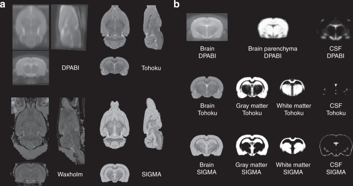
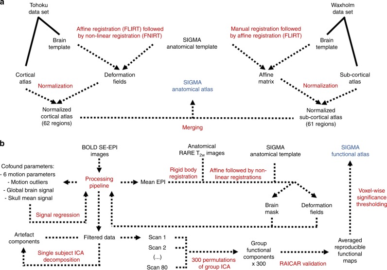
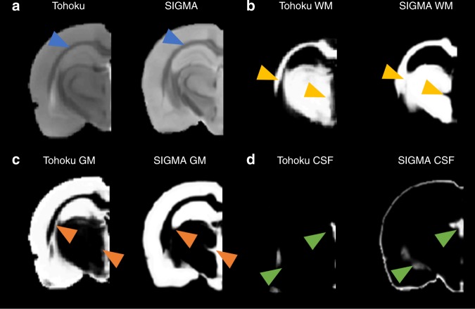
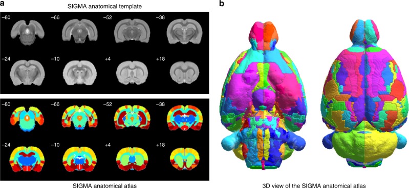
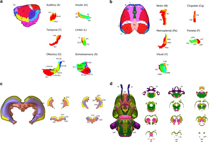
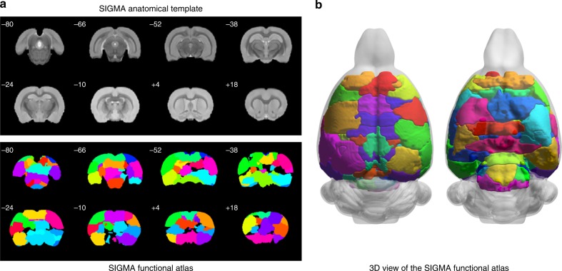
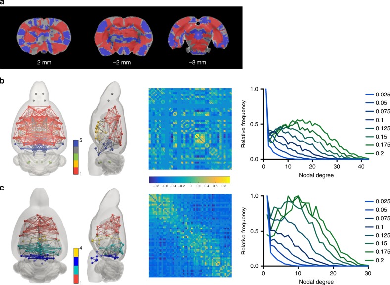
Preclinical imaging studies offer a unique access to the rat brain, allowing investigations that go beyond what is possible in human studies. Unfortunately, these techniques still suffer from a lack of dedicated and standardized neuroimaging tools, namely brain templates and descriptive atlases. Here, we present two rat brain MRI templates and their associated gray matter, white matter and cerebrospinal fluid probability maps, generated from ex vivo [Formula: see text]-weighted images (90 µm isotropic resolution) and in vivo T-weighted images (150 µm isotropic resolution). In association with these templates, we also provide both anatomical and functional 3D brain atlases, respectively derived from the merging of the Waxholm and Tohoku atlases, and analysis of resting-state functional MRI data. Finally, we propose a complete set of preclinical MRI reference resources, compatible with common neuroimaging software, for the investigation of rat brain structures and functions.
临床前成像研究为研究大鼠大脑提供了独特的途径,使我们能够进行一些在人体研究中无法进行的研究。不幸的是,这些技术仍然缺乏专用和标准化的神经影像学工具,即大脑模板和描述性图谱。在这里,我们展示了两个大鼠脑 MRI 模板及其相关的灰质、白质和脑脊液概率图,这些模板是由离体[公式:见正文]-加权图像(90µm 各向同性分辨率)和体内 T 加权图像(150µm 各向同性分辨率)生成的。与这些模板相关联,我们还分别提供了基于 Waxholm 和 Tohoku 图谱融合以及静息态功能 MRI 数据分析的解剖学和功能 3D 大脑图谱。最后,我们提出了一套完整的临床前 MRI 参考资源,与常见的神经影像学软件兼容,用于研究大鼠脑结构和功能。