Advanced Institute of Aging Science, Chonnam National University, Gwangju, 61186, Republic of Korea.
Department of Psychiatry, Massachusetts General Hospital and Harvard Medical School, Boston, 02129, USA.
Sci Rep. 2020 Jul 2;10(1):10912. doi: 10.1038/s41598-020-67873-y.
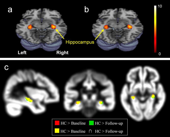
The efficacy of donepezil is well known for improving the cognitive performance in patients with mild cognitive impairment (MCI) and Alzheimer's disease (AD). Most of the recent neuroimaging studies focusing on the brain morphometry have dealt with the targeted brain structures, and thus it remains unknown how donepezil treatment influences the volume change over the whole brain areas including the cortical and subcortical regions and hippocampal subfields in particular. This study aimed to evaluate overall gray matter (GM) volume changes after donepezil treatment in MCI, which is a prodromal phase of AD, using voxel-based morphometry. Patients with MCI underwent the magnetic resonance imaging (MRI) before and after 6-month donepezil treatment. The cognitive function for MCI was evaluated using the questionnaires of the Korean version of the mini-mental state examination (K-MMSE) and Alzheimer's disease assessment scale-cognitive subscale (ADAS-Cog). Compared with healthy controls, patients with MCI showed significantly lower GM volumes in the hippocampus and its subfields, specifically in the right subiculum and left cornu ammonis (CA3). The average scores of K-MMSE in patients with MCI improved by 8% after donepezil treatment. Treated patients showed significantly higher GM volumes in the putamen, globus pailldus, and inferior frontal gyrus after donepezil treatment (p < 0.001). However, whole hippocampal volume in the patients decreased by 0.6% after 6-month treatment, and the rate of volume change in the left hippocampus was negatively correlated with the period of treatment. These findings will be useful for screening and tracking MCI, as well as understanding of the pathogenesis of MCI in connection with brain morphometric change.
多奈哌齐在改善轻度认知障碍(MCI)和阿尔茨海默病(AD)患者的认知表现方面的疗效众所周知。大多数最近关注脑形态计量学的神经影像学研究都针对靶向脑结构,因此尚不清楚多奈哌齐治疗如何影响包括皮质和皮质下区域以及海马亚区在内的整个大脑区域的体积变化。本研究旨在使用基于体素的形态计量学评估多奈哌齐治疗 MCI(AD 的前驱期)后总体灰质(GM)体积的变化。MCI 患者在多奈哌齐治疗前后接受磁共振成像(MRI)检查。使用韩国版简易精神状态检查表(K-MMSE)和阿尔茨海默病评估量表认知分量表(ADAS-Cog)评估 MCI 的认知功能。与健康对照组相比,MCI 患者的海马及其亚区,特别是右侧下托和左侧角回的 GM 体积明显较低。多奈哌齐治疗后,MCI 患者的 K-MMSE 平均评分提高了 8%。治疗组患者在治疗后纹状体、苍白球和额下回的 GM 体积明显增加(p<0.001)。然而,经过 6 个月的治疗,患者的整个海马体积减少了 0.6%,左侧海马的体积变化率与治疗期呈负相关。这些发现对于 MCI 的筛查和跟踪以及理解与脑形态计量学变化相关的 MCI 发病机制将非常有用。