• 文献检索
  • 文档翻译
  • 深度研究
  • 学术资讯
  • Suppr Zotero 插件Zotero 插件
  • 邀请有礼
  • 套餐&价格
  • 历史记录
应用&插件
Suppr Zotero 插件Zotero 插件浏览器插件Mac 客户端Windows 客户端微信小程序
定价
高级版会员购买积分包购买API积分包
服务
文献检索文档翻译深度研究API 文档MCP 服务
关于我们
关于 Suppr公司介绍联系我们用户协议隐私条款
关注我们

Suppr 超能文献

核心技术专利:CN118964589B侵权必究
粤ICP备2023148730 号-1Suppr @ 2026

文献检索

告别复杂PubMed语法,用中文像聊天一样搜索,搜遍4000万医学文献。AI智能推荐,让科研检索更轻松。

立即免费搜索

文件翻译

保留排版,准确专业,支持PDF/Word/PPT等文件格式,支持 12+语言互译。

免费翻译文档

深度研究

AI帮你快速写综述,25分钟生成高质量综述,智能提取关键信息,辅助科研写作。

立即免费体验

应用肺部超声鉴别 2019 年冠状病毒病(COVID-19)肺炎与社区获得性肺炎。

Use of Lung Ultrasound to Differentiate Coronavirus Disease 2019 (COVID-19) Pneumonia From Community-Acquired Pneumonia.

机构信息

Department of Critical Care Medicine, Second Affiliated Hospital of Fujian Medical University, Fujian, China; Department of Infectious Disease, Wuhan Jinyintan Hospital, Wuhan, China.

Collaborative Innovation Center for Maternal and Infant Health Service Application Technology of Education Ministry, Quanzhou Medical College, Fujian, China; Department of Ultrasound Medicine, Second Affiliated Hospital of Fujian Medical University, Fujian, China.

出版信息

Ultrasound Med Biol. 2020 Oct;46(10):2651-2658. doi: 10.1016/j.ultrasmedbio.2020.05.006. Epub 2020 Jun 5.

DOI:10.1016/j.ultrasmedbio.2020.05.006
PMID:32622684
原文链接:
https://pmc.ncbi.nlm.nih.gov/articles/PMC7274602/
Abstract

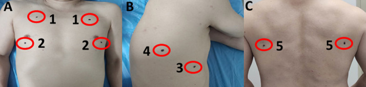
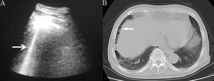
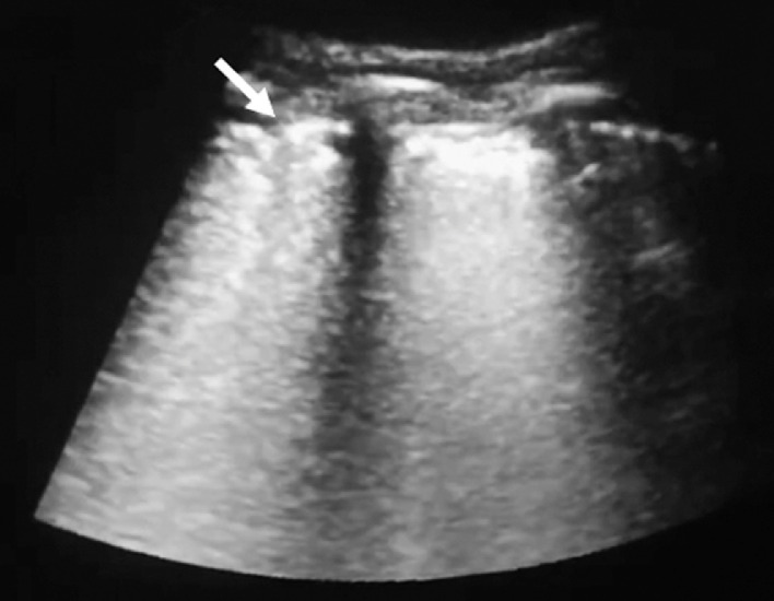
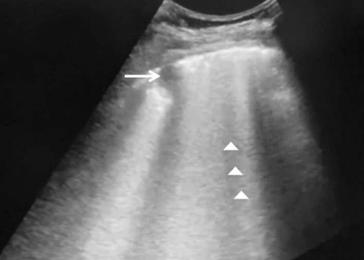
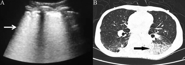
To investigate the feasibility of lung ultrasound in evaluating coronavirus disease 2019 (COVID-19) and distinguish the sonographic features between COVID-19 and community-acquired pneumonia (CAP), a total of 12 COVID-19 patients and 20 CAP patients were selected and underwent lung ultrasound. The modified Buda scoring system for interstitial lung disease was used to evaluate the severity and treatment effect of COVID-19 on ultrasonography. The differences between modified lung ultrasound (MLUS) score and high-resolution computed tomography (HRCT) Warrick score were analyzed to evaluate their correlation. COVID-19 showed the following sonographic features: thickening (12/12), blurred (9/12), discontinuous (6/12) pleural line; rocket sign (4/12), partially diffused B-line (12/12), completely diffused B-line (10/12), waterfall sign (4/12); C-line sign (5/12); pleural effusion (1/12) and pulmonary balloon (Am line, 1/12). The last two features were rarely seen. Differences of ultrasonic features, including lesion range, lung signs and pneumonia-related complications, between COVID-19 and CAP were statistically significant (p˂ 0.05 or 0.001). MLUS scores (p = 0.006) and HRCT Warrick scores (p = 0.015) increased as the severity of COVID-19 increased. The differences between moderate (29.00 [25.75-37.50]) and severe (43.00 [38.75-47.25]) (p = 0.022) or between moderate and critical (47.50 [44.25-50.00]) (p = 0.002) type COVID-19 were statistically significant, compared with those between severe and critical types. Correlation between MLUS scores and HRCT Warrick scores was positive (r = 0.54, p = 0.048). MLUS scores (Z = 2.61, p = 0.009) and HRCT Warrick scores (Z = 2.63, p = 0.009) of five severe or critical COVID-19 patients significantly decreased as their conditions improved after treatment. The differences of sonographic features between COVID-19 and CAP patients were notable. The MLUS scoring system could be used to evaluate the severity and treatment effect of COVID-19.

摘要

目的 探讨肺部超声在评估 2019 冠状病毒病(COVID-19)中的可行性,并区分 COVID-19 与社区获得性肺炎(CAP)的声像特征。选择了 12 例 COVID-19 患者和 20 例 CAP 患者进行肺部超声检查。采用改良的间质性肺疾病 Buda 评分系统评估 COVID-19 超声严重程度和治疗效果。分析改良肺部超声(MLUS)评分与高分辨率 CT(HRCT)Warrick 评分的差异,以评估其相关性。结果 COVID-19 的超声特征如下:增厚(12/12)、模糊(9/12)、不连续(6/12)胸膜线;火箭征(4/12)、部分弥漫性 B 线(12/12)、完全弥漫性 B 线(10/12)、瀑布征(4/12);C 线征(5/12);胸腔积液(1/12)和肺气囊(Am 线,1/12)。后两种特征很少见。COVID-19 与 CAP 之间的超声特征差异,包括病变范围、肺部征象和肺炎相关并发症,具有统计学意义(p˂0.05 或 0.001)。MLUS 评分(p=0.006)和 HRCT Warrick 评分(p=0.015)随 COVID-19 严重程度的增加而升高。中度(29.00[25.75-37.50])和重度(43.00[38.75-47.25])(p=0.022)或中度和危重症(47.50[44.25-50.00])(p=0.002)COVID-19 类型之间的差异有统计学意义,而重度和危重症 COVID-19 类型之间的差异无统计学意义。MLUS 评分与 HRCT Warrick 评分呈正相关(r=0.54,p=0.048)。治疗后,5 例重症或危重症 COVID-19 患者的 MLUS 评分(Z=2.61,p=0.009)和 HRCT Warrick 评分(Z=2.63,p=0.009)显著降低,病情改善。COVID-19 与 CAP 患者的超声特征差异显著。MLUS 评分系统可用于评估 COVID-19 的严重程度和治疗效果。

https://cdn.ncbi.nlm.nih.gov/pmc/blobs/306a/7274602/d76de23a76e8/gr5_lrg.jpg
https://cdn.ncbi.nlm.nih.gov/pmc/blobs/306a/7274602/c49b65bb8c14/gr1_lrg.jpg
https://cdn.ncbi.nlm.nih.gov/pmc/blobs/306a/7274602/7f58219f00dd/gr2_lrg.jpg
https://cdn.ncbi.nlm.nih.gov/pmc/blobs/306a/7274602/d4ecf6e91b69/gr3_lrg.jpg
https://cdn.ncbi.nlm.nih.gov/pmc/blobs/306a/7274602/5117f45e649d/gr4_lrg.jpg
https://cdn.ncbi.nlm.nih.gov/pmc/blobs/306a/7274602/d76de23a76e8/gr5_lrg.jpg

相似文献

1
Use of Lung Ultrasound to Differentiate Coronavirus Disease 2019 (COVID-19) Pneumonia From Community-Acquired Pneumonia.应用肺部超声鉴别 2019 年冠状病毒病(COVID-19)肺炎与社区获得性肺炎。
Ultrasound Med Biol. 2020 Oct;46(10):2651-2658. doi: 10.1016/j.ultrasmedbio.2020.05.006. Epub 2020 Jun 5.
2
Differential diagnosis of coronavirus disease 2019 from community-acquired-pneumonia by computed tomography scan and follow-up.通过计算机断层扫描和随访对 2019 年冠状病毒病与社区获得性肺炎进行鉴别诊断。
Infect Dis Poverty. 2020 Aug 26;9(1):118. doi: 10.1186/s40249-020-00737-9.
3
Similarities and Differences of Early Pulmonary CT Features of Pneumonia Caused by SARS-CoV-2, SARS-CoV and MERS-CoV: Comparison Based on a Systemic Review.新型冠状病毒、严重急性呼吸综合征冠状病毒和中东呼吸综合征冠状病毒所致肺炎早期肺部CT特征的异同:基于系统评价的比较
Chin Med Sci J. 2020 Sep 30;35(3):254-261. doi: 10.24920/003727.
4
[Clinical features and high resolution CT imaging evolution of coronavirus disease 2019].新型冠状病毒肺炎的临床特征及高分辨率CT影像学演变
Zhonghua Jie He He Hu Xi Za Zhi. 2020 Jun 12;43(6):509-515. doi: 10.3760/cma.j.cn112147-20200214-00094.
5
Analysis of clinical features and imaging signs of COVID-19 with the assistance of artificial intelligence.人工智能辅助分析 COVID-19 的临床特征和影像征象。
Eur Rev Med Pharmacol Sci. 2020 Aug;24(15):8210-8218. doi: 10.26355/eurrev_202008_22510.
6
Lung Ultrasound Score in Evaluating the Severity of Coronavirus Disease 2019 (COVID-19) Pneumonia.肺部超声评分在评估 2019 年冠状病毒病(COVID-19)肺炎严重程度中的作用。
Ultrasound Med Biol. 2020 Nov;46(11):2938-2944. doi: 10.1016/j.ultrasmedbio.2020.07.024. Epub 2020 Jul 24.
7
CT imaging features of COVID-19 pneumonia: initial experience from Turkey.土耳其的 COVID-19 肺炎 CT 影像学特征:初步经验。
Diagn Interv Radiol. 2020 Jul;26(4):308-314. doi: 10.5152/dir.2020.20307.
8
Lung Ultrasound May Support Diagnosis and Monitoring of COVID-19 Pneumonia.肺部超声检查可能有助于 COVID-19 肺炎的诊断和监测。
Ultrasound Med Biol. 2020 Nov;46(11):2908-2917. doi: 10.1016/j.ultrasmedbio.2020.07.018. Epub 2020 Jul 20.
9
Lung ultrasound for early diagnosis and severity assessment of pneumonia in patients with coronavirus disease 2019.肺部超声在 2019 年冠状病毒病患者肺炎的早期诊断和严重程度评估中的应用。
Korean J Intern Med. 2020 Jul;35(4):771-781. doi: 10.3904/kjim.2020.180. Epub 2020 Jul 1.
10
A Comparison of Clinical and Chest CT Findings in Patients With Influenza A (H1N1) Virus Infection and Coronavirus Disease (COVID-19).甲型流感病毒(H1N1)感染与新型冠状病毒肺炎(COVID-19)患者的临床和胸部 CT 表现比较。
AJR Am J Roentgenol. 2020 Nov;215(5):1065-1071. doi: 10.2214/AJR.20.23214. Epub 2020 May 26.

引用本文的文献

1
Pulmonary ultrasound in COVID-19 and non-COVID-19 pneumonia in South Africa: An observational study.南非新冠肺炎与非新冠肺炎肺炎的肺部超声:一项观察性研究。
Afr J Thorac Crit Care Med. 2025 Mar 28;31(1):e1887. doi: 10.7196/AJTCCM.2025.v31i1.1887. eCollection 2025.
2
Distinguishing types and severity of pediatric pneumonia using modified lung ultrasound score.使用改良肺部超声评分法区分小儿肺炎的类型和严重程度。
Front Pediatr. 2024 Aug 5;12:1411365. doi: 10.3389/fped.2024.1411365. eCollection 2024.
3
Current Diagnostic Techniques for Pneumonia: A Scoping Review.

本文引用的文献

1
The Epidemiological Characteristics of an Outbreak of 2019 Novel Coronavirus Diseases (COVID-19) - China, 2020.2019新型冠状病毒病(COVID-19)疫情的流行病学特征 - 中国,2020年
China CDC Wkly. 2020 Feb 21;2(8):113-122.
2
Pathological findings of COVID-19 associated with acute respiratory distress syndrome.与急性呼吸窘迫综合征相关的新型冠状病毒肺炎的病理表现
Lancet Respir Med. 2020 Apr;8(4):420-422. doi: 10.1016/S2213-2600(20)30076-X. Epub 2020 Feb 18.
3
A rapid advice guideline for the diagnosis and treatment of 2019 novel coronavirus (2019-nCoV) infected pneumonia (standard version).
肺炎的当前诊断技术:范围综述。
Sensors (Basel). 2024 Jul 1;24(13):4291. doi: 10.3390/s24134291.
4
Lung Ultrasound and High-Resolution Computed Tomography in Suspected COVID-19 Patients Admitted to the Emergency Department: A Comparison.急诊科收治的疑似新型冠状病毒肺炎患者的肺部超声与高分辨率计算机断层扫描:一项比较研究
J Diagn Med Sonogr. 2023 Jan 29;39(4):332-346. doi: 10.1177/87564793221147496. eCollection 2023 Jul-Aug.
5
Point-of-care ultrasound use in COVID-19: a narrative review.即时超声在2019冠状病毒病中的应用:一项叙述性综述。
Ann Transl Med. 2024 Feb 1;12(1):13. doi: 10.21037/atm-23-1403. Epub 2023 Jul 13.
6
Lung Ultrasound Score in COVID-19 Patients Correlates with PO/FiO, Intubation Rates, and Mortality.新冠肺炎患者的肺部超声评分与 PO/FiO、插管率和死亡率相关。
West J Emerg Med. 2024 Jan;25(1):28-39. doi: 10.5811/westjem.59975.
7
Evaluation of extravascular lung water and cardiac function in normal vaginal delivery by intrapartum bedside ultrasound.产时床边超声评估正常阴道分娩产妇的肺血管外水和心功能。
BMC Pregnancy Childbirth. 2024 Jan 2;24(1):13. doi: 10.1186/s12884-023-06201-4.
8
Italian Society of Anesthesia, Analgesia, Resuscitation, and Intensive Care expert consensus statement on the use of lung ultrasound in critically ill patients with coronavirus disease 2019 (ITACO).意大利麻醉、镇痛、复苏与重症监护学会关于2019冠状病毒病(COVID-19)重症患者肺部超声应用的专家共识声明(ITACO)。
J Anesth Analg Crit Care. 2021 Nov 24;1(1):16. doi: 10.1186/s44158-021-00015-6.
9
A study of the diagnostic value of a modified transthoracic lung ultrasound scoring method in interstitial lung disease.改良经胸肺超声评分法在间质性肺疾病中的诊断价值研究
Quant Imaging Med Surg. 2023 Feb 1;13(2):946-956. doi: 10.21037/qims-22-153. Epub 2023 Jan 14.
10
Egyptian Consensus on the Role of Lung Ultrasonography During the Coronavirus Disease 2019 Pandemic.埃及关于肺部超声在2019年冠状病毒病大流行期间作用的共识
Infect Drug Resist. 2022 Apr 20;15:1995-2013. doi: 10.2147/IDR.S353283. eCollection 2022.
新型冠状病毒(2019-nCoV)感染的肺炎快速诊治指南(标准版)。
Mil Med Res. 2020 Feb 6;7(1):4. doi: 10.1186/s40779-020-0233-6.
4
Early Transmission Dynamics in Wuhan, China, of Novel Coronavirus-Infected Pneumonia.新型冠状病毒感染肺炎在中国武汉的早期传播动力学。
N Engl J Med. 2020 Mar 26;382(13):1199-1207. doi: 10.1056/NEJMoa2001316. Epub 2020 Jan 29.
5
Point-of-Care Ultrasound-A New Option for Early Quantitative Assessment of Pulmonary Edema.即时超声——一种早期定量评估肺水肿的新选择。
Ultrasound Med Biol. 2020 Jan;46(1):1-10. doi: 10.1016/j.ultrasmedbio.2019.08.008. Epub 2019 Sep 28.
6
Diagnosis and Treatment of Adults with Community-acquired Pneumonia. An Official Clinical Practice Guideline of the American Thoracic Society and Infectious Diseases Society of America.成人社区获得性肺炎诊断和治疗。美国胸科学会和美国传染病学会的官方临床实践指南。
Am J Respir Crit Care Med. 2019 Oct 1;200(7):e45-e67. doi: 10.1164/rccm.201908-1581ST.
7
Prospective observational study of point-of-care ultrasound for diagnosing pneumonia.以床旁超声诊断肺炎的前瞻性观察性研究。
Arch Dis Child. 2019 Jan;104(1):12-18. doi: 10.1136/archdischild-2017-314496. Epub 2018 Jun 7.
8
Systematic review and meta-analysis for the use of ultrasound versus radiology in diagnosing of pneumonia.超声与放射学在肺炎诊断中应用的系统评价与荟萃分析
Crit Ultrasound J. 2017 Dec;9(1):6. doi: 10.1186/s13089-017-0059-y. Epub 2017 Feb 27.
9
Lung Ultrasound for Early Diagnosis of Ventilator-Associated Pneumonia.肺部超声在呼吸机相关性肺炎早期诊断中的应用。
Chest. 2016 Apr;149(4):969-80. doi: 10.1016/j.chest.2015.12.012. Epub 2015 Dec 22.
10
Lung Ultrasonography in the Evaluation of Interstitial Lung Disease in Systemic Connective Tissue Diseases: Criteria and Severity of Pulmonary Fibrosis - Analysis of 52 Patients.肺部超声在系统性结缔组织病间质性肺疾病评估中的应用:肺纤维化的标准与严重程度——52例患者分析
Ultraschall Med. 2016 Aug;37(4):379-85. doi: 10.1055/s-0041-110590. Epub 2015 Dec 29.