Gool Jari K, van der Werf Ysbrand D, Lammers Gert Jan, Fronczek Rolf
Department of Anatomy and Neurosciences, Amsterdam UMC, Location VUmc, De Boelelaan 1108, 1081HZ Amsterdam, Noord-Holland, The Netherlands.
Sleep-Wake Centre SEIN, Achterweg 2, 2103SW Heemstede, Noord-Holland, The Netherlands.
Brain Sci. 2020 Jul 1;10(7):419. doi: 10.3390/brainsci10070419.
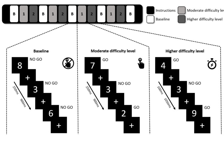
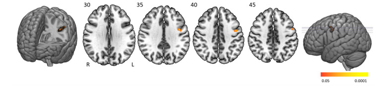
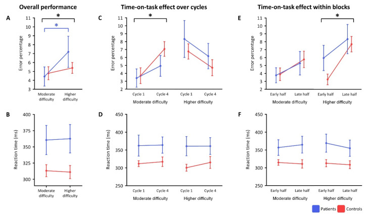
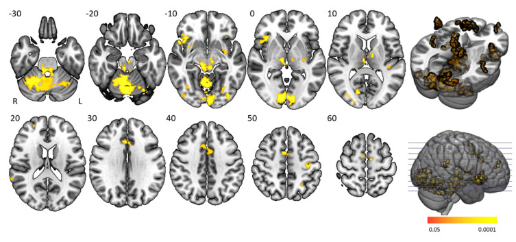
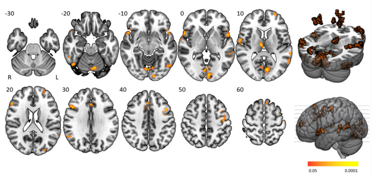
Vigilance complaints often occur in people with narcolepsy type 1 and severely impair effective daytime functioning. We tested the feasibility of a three-level sustained attention to response task (SART) paradigm within a magnetic resonance imaging (MRI) environment to understand brain architecture underlying vigilance regulation in individuals with narcolepsy type 1. Twelve medication-free people with narcolepsy type 1 and 11 matched controls were included. The SART included four repetitions of a baseline block and two difficulty levels requiring moderate and high vigilance. Outcome measures were between and within-group performance indices on error rates and reaction times, and functional MRI (fMRI) parameters: mean activity during the task and between-group activity differences across the three conditions and related to changes in activation over time (time-on-task) and error-related activity. Patients-but not controls-made significantly more mistakes with increasing difficulty. The modified SART is a feasible MRI vigilance task showing similar task-positive brain activity in both groups within the cingulo-opercular, frontoparietal, arousal, motor, and visual networks. During blocks of higher vigilance demand, patients had significantly lower activation in these regions than controls. Patients had lower error-related activity in the left pre- and postcentral gyrus. The time-on-task activity differences between groups suggest that those with narcolepsy are insufficiently capable of activating attention- and arousal-related regions when transitioning from attention initiation to stable attention, specifically when vigilance demand is high. They also show lower inhibitory motor activity in relation to errors, suggesting impaired executive functioning.
警觉性抱怨在1型发作性睡病患者中经常出现,并严重损害白天的有效功能。我们在磁共振成像(MRI)环境中测试了三级持续注意力反应任务(SART)范式的可行性,以了解1型发作性睡病患者警觉性调节的脑结构。研究纳入了12名未服用药物的1型发作性睡病患者和11名匹配的对照组。SART包括基线阶段的四次重复以及两个需要中等和高度警觉性的难度级别。结果指标包括组间和组内关于错误率和反应时间的表现指数,以及功能磁共振成像(fMRI)参数:任务期间的平均活动、三种条件下的组间活动差异以及与激活随时间变化(任务持续时间)和错误相关活动的变化。随着难度增加,患者(而非对照组)出现的错误显著增多。改良后的SART是一项可行的MRI警觉性任务,在扣带回 - 脑岛、额顶叶、觉醒、运动和视觉网络中,两组的任务阳性脑活动相似。在更高警觉性需求阶段,患者在这些区域的激活明显低于对照组。患者在左中央前回和中央后回的错误相关活动较低。组间任务持续时间的活动差异表明,发作性睡病患者在从注意力启动过渡到稳定注意力时,尤其是在警觉性需求较高时,激活注意力和觉醒相关区域的能力不足。他们在与错误相关的抑制性运动活动方面也较低,表明执行功能受损。