Cognitive Neuroscience Laboratory, German Primate Center - Leibniz Institute for Primate Research, 37077, Göttingen, Germany.
Primate Genetics Laboratory, German Primate Center - Leibniz Institute for Primate Research, 37077, Göttingen, Germany.
Sci Rep. 2020 Jul 6;10(1):11051. doi: 10.1038/s41598-020-67752-6.
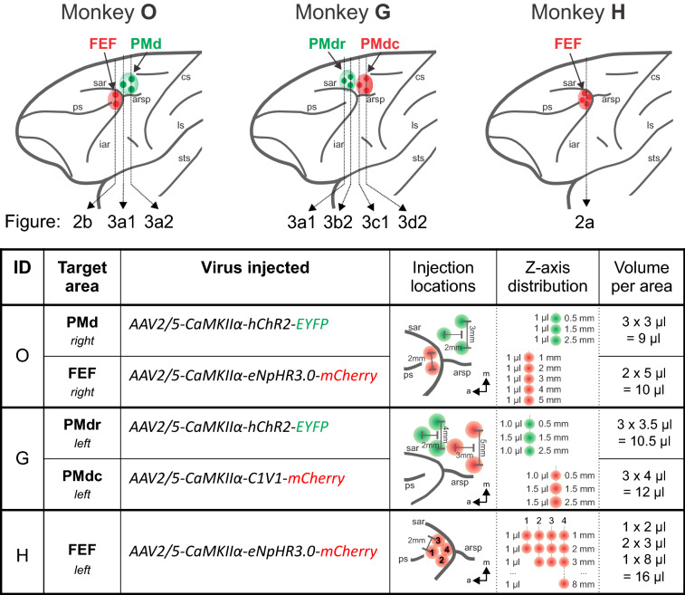
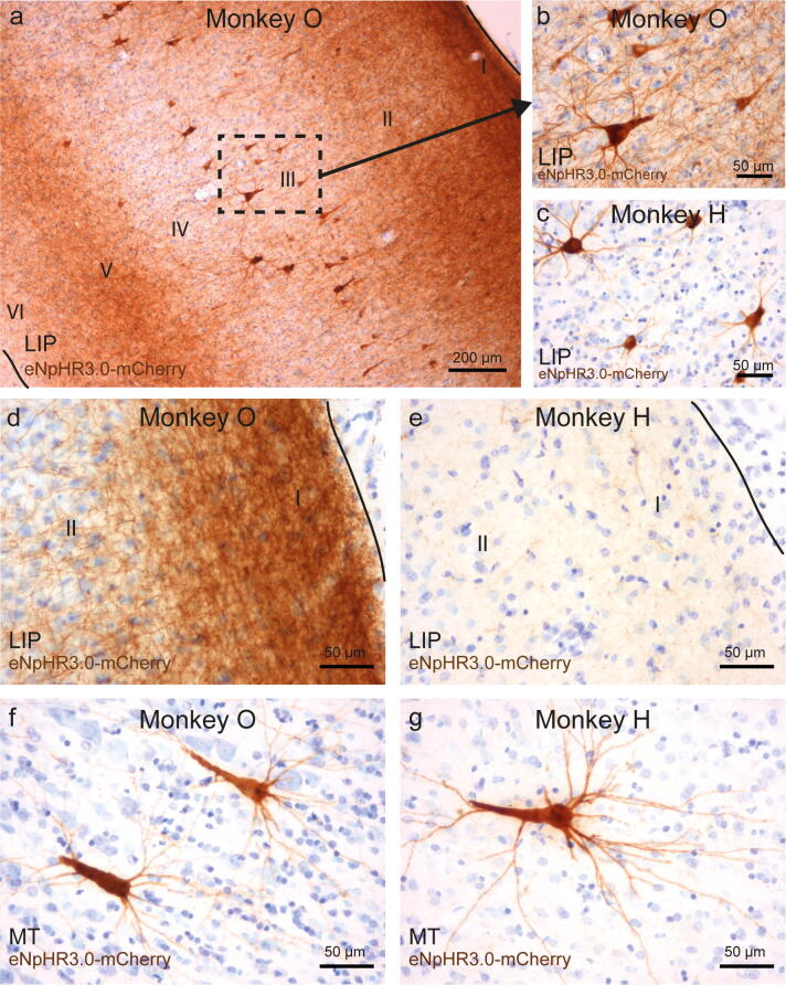
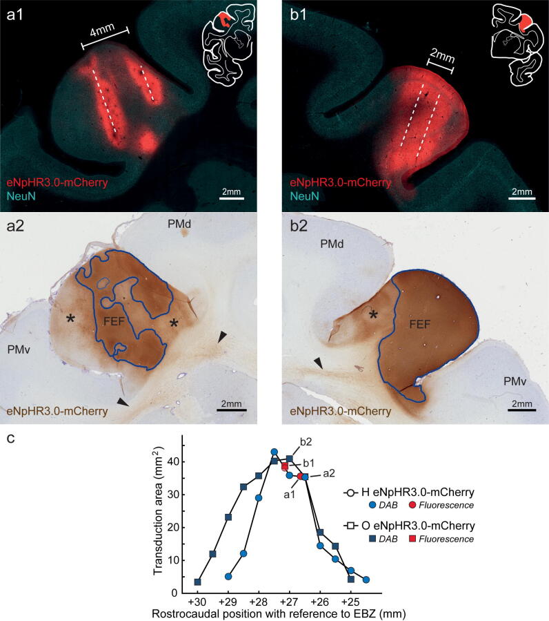
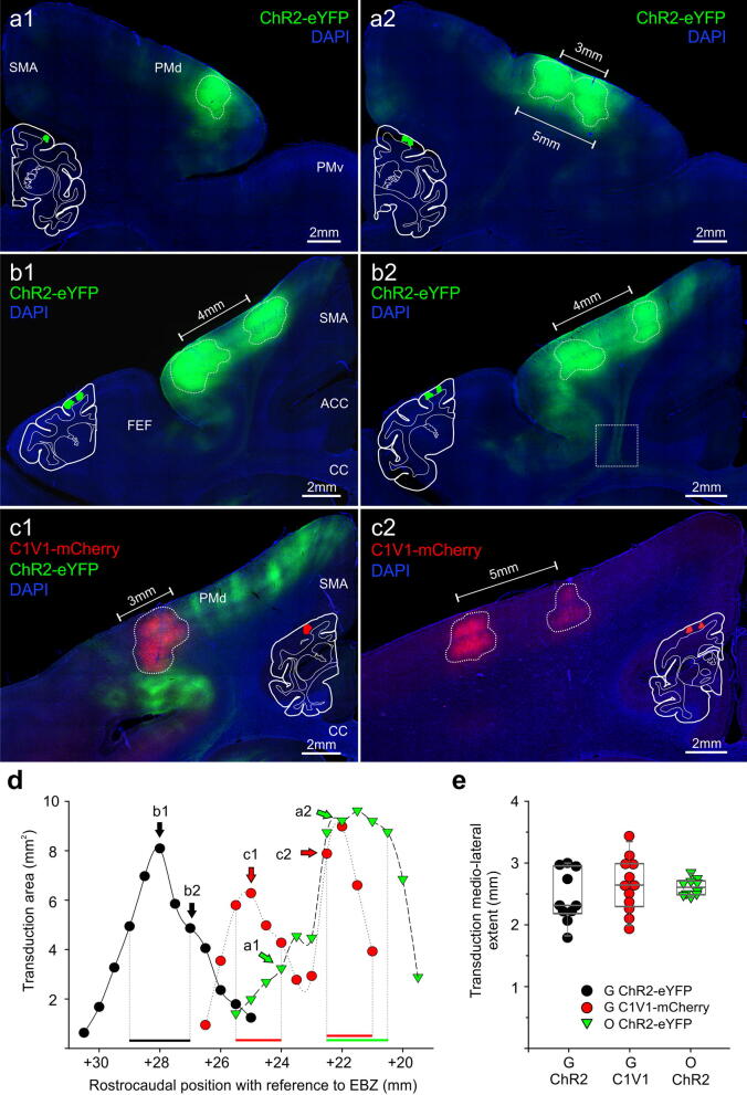
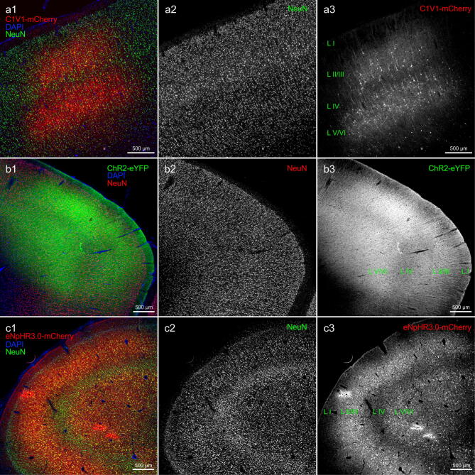
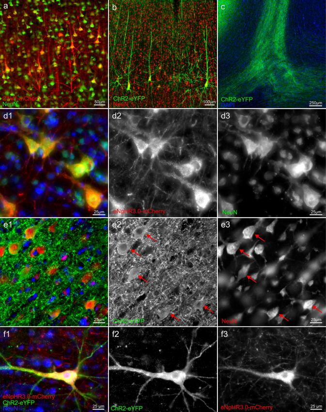
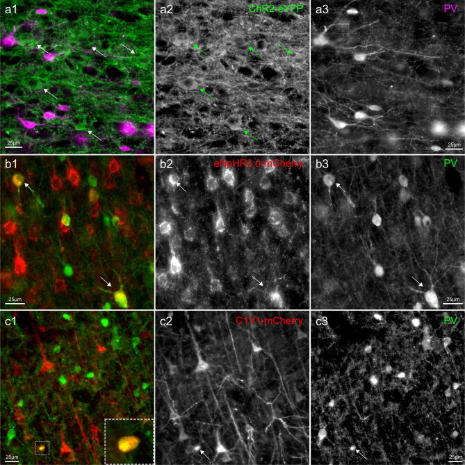
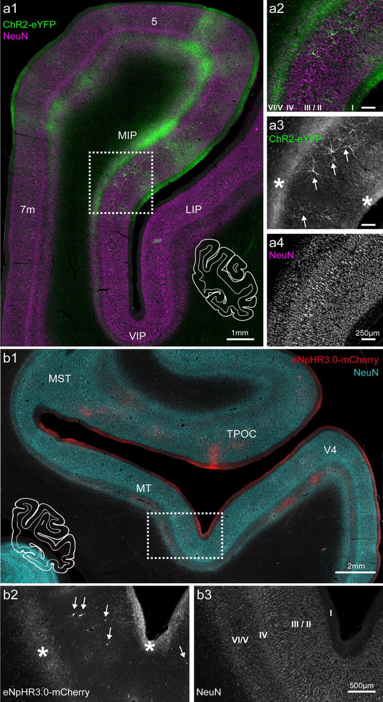
Optogenetics offers unprecedented possibilities to investigate cortical networks. Yet, the number of successful optogenetic applications in non-human primates is still low, and the consequences of opsin expression in the primate brain are not well documented. We assessed histologically if we can target cerebrocortical networks with three common optogenetic constructs (AAV2/5-CaMKIIα-eNpHR3.0-mCherry, -ChR2-eYFP, -C1V1-mCherry). The frontal eye field or the dorsal premotor area of rhesus macaques were virally injected, and the resulting transduction spread, expression specificity, and opsin trafficking into axons projecting to parietal and visual areas were examined. After variable periods (2-24 months), expression was robust for all constructs at the injection sites. The CaMKIIα promoter driven-expression was predominant, but not exclusive, in excitatory neurons. In the case of eNpHR3.0-mCherry and ChR2-eYFP, opsins were present in axonal projections to target areas, in which sparse, retrogradely transduced neurons could also be found. Finally, the intracellular distribution of opsins differed: ChR2-eYFP had almost exclusive membrane localization, while eNpHR3.0-mCherry and C1V1-mCherry showed additional intracellular accumulations, which might affect neuronal survival in the long-term. Results indicate that all three constructs can be used for local neuronal modulation, but axonal stimulation and long-term use require additional considerations of construct selection and verification.
光遗传学为研究皮质网络提供了前所未有的可能性。然而,在非人类灵长类动物中成功应用光遗传学的数量仍然很低,并且在灵长类动物大脑中表达光感受蛋白的后果也没有得到很好的记录。我们通过组织学评估了三种常见的光遗传学构建体(AAV2/5-CaMKIIα-eNpHR3.0-mCherry、-ChR2-eYFP、-C1V1-mCherry)是否可以靶向脑皮质网络。我们通过病毒注射恒河猴的额眼区或背侧运动前区,并检查由此产生的转导扩散、表达特异性以及向投射到顶叶和视觉区域的轴突中的光感受蛋白运输情况。在不同的时间段(2-24 个月)后,所有构建体在注射部位的表达都很稳定。CaMKIIα 启动子驱动的表达在兴奋性神经元中占主导地位,但不是唯一的。在 eNpHR3.0-mCherry 和 ChR2-eYFP 的情况下,光感受蛋白存在于轴突投射到靶区中,在这些区域中也可以发现稀疏的逆行转导神经元。最后,光感受蛋白的细胞内分布不同:ChR2-eYFP 几乎完全定位于膜上,而 eNpHR3.0-mCherry 和 C1V1-mCherry 则显示出额外的细胞内积累,这可能会影响神经元的长期存活。结果表明,所有三种构建体都可用于局部神经元调节,但轴突刺激和长期使用需要进一步考虑构建体的选择和验证。